ᐅ SummaryArea 3a: Part of primary sensory area. along with area 2, is known to receive sensory information from deep body tissues. This area is also involved in the burning, chronic pain sensations that originate from deep somatic tissue. This suggests area 3a has relevance in many of the chronic pain ailments observed in the clinical setting. Area 3a is also involved in proprioception. ᐅ Where is it?Area 3a is found in the depth of the central sulcus. It follows this sulcus up to the midline but does not fold onto the medial face of the hemisphere. ᐅ What are its borders?Area 3a borders area 2 along its long anterior border and area 3b along its posterior border. Inferiorly, it borders area 23 and superiorly it borders area 5m. It has a small area of posterior contact with area 2 near the Sylvian fissure. ᐅ What are its borders?Area 3a demonstrates functional connectivity areas 1, 2, 4, 3b in the motor and sensory strips, areas SCEF, 55b, 6d, 6v, and 6mp in the premotor regions, areas 24dd, 24dv, 5m, and 5L in the middle cingulate regions, areas 43, OP1, OP2-3, OP4, IG, and FOP2 in the superior insula opercular regions, areas PoI2, 52, A4, A5, RI, A1, MBelt, PBelt, LBelt, TA2, and STV in the lower opercula and Heschl's gyrus regions, areas PFcm, 7AL, and 7PC, in the parietal lobe, areas V2, V3, and V4 in the medial occipital lobe, areas V6, V6a, V3a, and V7 in the dorsal visual stream areas, area FFC of the ventral visual stream, and areas PH, TPOJ1, FST, V4t, MST, MT, and LO3 of the lateral occipital lobe. ᐅ What are its functional connections?Area 3a is structurally connected to the pyramidal tracts, thalamocortical projections, contralateral hemisphere and the parietal lobe. Connections to pyramidal tracts descend through the posterior limb of the internal capsule and cerebral peduncle to the brainstem. Thalamocortical tracts run medial to pyramidal projections to enter the thalamus. Contralateral connections course through the body of the corpus callosum to parcellations 3b and 4. Parietal projections are portions of the SLF and connect to PFm. Local short association fibers are connected with 4, 3b, 2, 1, and 6v. ᐅ What are its white matter connections?Area 3a, along with area 2, is known to receive sensory information from deep body tissues. This area is also involved in the burning, chronic pain sensations that originate from deep somatic tissue. This suggests area 3a has relevance in many of the chronic pain ailments observed in the clinical setting. Area 3a is also involved in proprioception. |
|
A: lateral-medial
B: anterior-posterior
C: superior-inferior
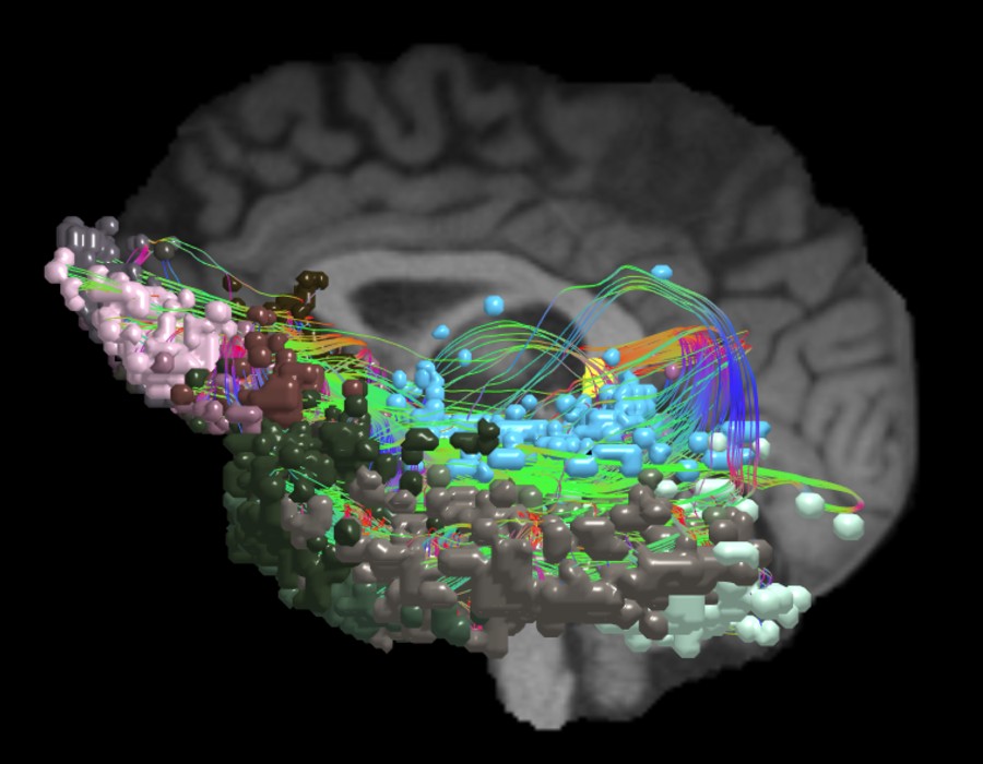
DTI image |


ᐅ SummaryArea 4: part of primary motor cortex. Known to produce fine motor movements of the distal forearm and fingers. Additionally, it functions in giving a muscle its tone and producing forceful muscle contractions. It is also thought to play a role in visual learning of motor-based skills in the early stages of life. ᐅ Where is it?Area 4 is found in the precentral gyrus. It is predominantly located on the posterior half of the gyrus, making up the anterior bank of the central sulcus. It widens medially to fill a larger proportion of the paracentral lobule, which is the leg motor region. The somatotopic organization of this area is well known, however it is worth noting that there is an area dedicated to the eye which is adjacent to the frontal eye fields. ᐅ What are its borders?Area 4 borders area 3a along its posterior convexity border. Its inferior end contains area 43. At its superior end, on the medial surface (i.e. the paracentral lobule), it borders area 5m posteriorly, area 44dd inferiorly, and area 6mp anteriorly. Its long anterior border contacts several areas, including (from superior to inferior): area 6d, FEF, area 55b, PEF, and area 6v, most of which contact it on the anterior half of the precentral gyrus. ᐅ What are its borders?Area 4 demonstrates functional connectivity to areas 1, 2, 3a, 3b in the sensory strip, areas SCEF, 55b, 6d, 6v, and 6mp in the premotor regions, areas 24dd, 24dv, 5m, and 5L in the middle cingulate regions, areas 43, OP1, OP2-3, OP4, IG, and FOP2 in the superior insula opercular regions, areas A4, A5, RI, PBelt, LBelt, TA2, and STV in the lower opercula and Heschl's gyrus regions, areas VIP, IPS1, LIPv and 7PC, in the parietal lobe, areas V2, V3, and V4 in the medial occipital lobe, areas V6, V6a, V3a, and V7 in the dorsal visual stream areas, area FFC of the ventral visual stream, and areas PH, TPOJ1, FST, V4t, MT, and LO3 of the lateral occipital lobe. ᐅ What are its functional connections?Area 4 is structurally connected to pyramidal tracts, the contralateral hemisphere and the parietal lobule. Connections to pyramidal tracts descend through the posterior limb of the internal capsule and cerebral peduncle to the brainstem. Contralateral connections course through the body of the corpus callosum to parcellations 4, 6ma and 6mp. Parietal projections are portions of the SLF and connect to PFm. Local short association fibers are connected with 3a, 3b, 2, 1, and 6v. ᐅ What are its white matter connections?Area 4 is known to produce fine motor movements of the distal forearm and fingers. Additionally, it functions in giving a muscle its tone and producing forceful muscle contractions. It is also thought to play a role in visual learning of motor-based skills in the early stages of life. |
|
A: lateral-medial
B: anterior-posterior
C: superior-inferior
DTI image |


ᐅ SummaryArea 46: part of the lateral frontal lobe. Plays a role in goal-directed higher-order cognitive processes. The mid dorsolateral prefrontal cortex, which includes areas 9-46 and 46, is also involved in the conscious, active control of planned behavior. ᐅ Where is it?Area 46 parallels area 9-46d along its slightly oblique course. It begins in the depths of the SFS posteriorly and its anterior extent spills onto the MFG. ᐅ What are its borders?Area 46 borders all three 9-46 hybrid regions, as well as several others, giving it a central place in the ventral prefrontal cortex. Its medial border is mainly area 9-46d and its lateral border is mainly area p9-46v, both of which roughly parallel it along its oblique course. It has an anterolateral border with IFSa, as IFSa rises onto the lateral bank of MFG to meet it. Its anterior limit is mainly with area a9-46v. Its posterior limit forms a wedge with areas 8AV and 8AD. ᐅ What are its borders?Area 46 demonstrates functional connectivity to areas a9-46v, p9-46v, and IFSa in the dorsolateral frontal lobe, areas SCEF, a32prime, p24prime, and a24prime in the medial frontal lobe, areas 6ma, 6a, and 6r in the premotor regions, area 11L in the orbitofrontal region areas FOP1, FOP3, FOP4, FOP5, 52 PFop, PFcm, 43 PoI2, PoI1, and MI in the insula-opercular region, area PHT in the temporal lobe, areas PF, PFt, PGp, AIP, MIP 7AL, 7PL, and LIPd IP2 and IP0 in the parietal lobe, and areas 23c, PCV, POS2, PCV, 7am, 7pm, 23c, and DVT in the medial parietal lobe. It is also connected to much of the occipital lobe, showing connectivity to V1, V2, V3, V4, V3a, and V6. ᐅ What are its functional connections?Area 46 is structurally connected to local parcellations and the contralateral hemisphere. Contralateral connections travel through the corpus callosum to end at 9-46d and p9-47v. There are abundant local short association bundles connecting with 9-46d, a9-46v, p9-46v, IFSp and IFSa. ᐅ What are its white matter connections?Area 46 plays a role in goal-directed higher-order cognitive processes. The mid-dorsolateral prefrontal cortex, which includes areas 9-46 and 46, is also involved in the conscious, active control of planned behavior. |
|
A: lateral-medial
B: anterior-posterior
C: superior-inferior
DTI image |


ᐅ SummaryArea A4 (auditory area 4): part of temporal hypotenuse regions. Parcellated from the auditory association cortex. There is evidence that this regions of the brain processes perceptual and conceptual acoustic sounds. ᐅ Where is it?Area A4 (auditory area 4) is on the superior face of the posterior half of the STG. It occupies this portion of the gyrus posterior to its junction with Heschl's gyrus. ᐅ What are its borders?Area A4 borders A5 laterally and PBelt medially. Its posterior borders are with PSL and STV. Its anterior border is with MBelt and TA2. ᐅ What are its borders?Area A4 demonstrates functional connectivity to areas 1, 2,3a and 3b in the sensory strip, area 4 in the motor strip, areas SCEF, FEF, 6mp, and 6v in the premotor regions, areas 5m, 5l, and 24dd in the medial frontal lobe, areas 43, PFcm, OP4, OP2-3 and OP1 in the superior insula opercular regions, areas STV, 52, RI, TA2, PI, MI, PBelt, MBelt, LBelt, A1, A5, PoI1, and PoI2 in the lower opercula and Heschl's gyrus regions, areas 7PC and PFop, in the lateral parietal lobe, areas V2, V3, and V4 in the medial occipital lobe, areas V6, V6a, V7, and V3a in the dorsal visual stream area, areas V8, FFC, VVC, and PIT in the ventral visual stream regions, and areas TPOJ1, TPOJ2, LO1, LO3, MT, MST, PH, and FST in the lateral occipital lobe ᐅ What are its functional connections?Area A4 is structurally connected to the arcuate/SLF and middle longitudinal fasciculus. Arcuate/SLF fibers wrap around the termination of the sylvian fissure from A4 to inferior frontal gyrus parcellations 45 and FOP5. The middle longitudinal fasciculus projects posterior from A4 to run just lateral to the lateral ventricle ending at occipital and parietal lobe parcellations V2, V3, V6, V7, MIP, LIPd, LIPv and IP1. Local short association bundles connect with A5, PFop, RI, STV, LBelt, MBelt, PBelt and PFcm ᐅ What are its white matter connections?Area A4 is a newly described area of the brain parcellated from the auditory association cortex. There is evidence that this region of the brain processes perceptual and conceptual acoustic sounds. Area A4 was differentiated from area TA2 based on differences in fMRI activity during arithmetic and auditory story tasks. |
|
A: lateral-medial
B: anterior-posterior
C: superior-inferior
DTI image |


ᐅ SummaryArea AIP (anterior intraparietal): part of the lateral parietal lobe regions. Involved in grasping activity as well as object recognition. Neurons in this part of the cortex are oriented for grip and hand shape, and the inferior temporal cortex provides input related to object information. Receives input from the ventral and dorsolateral visual streams. Plays a role in shaping the hand for grasping activity. Beyond grasping action, is involved in tactile shape-processing and understanding orientation in space. ᐅ Where is it?Area AIP (Anterior intraparietal) is found on the superior bank of the intraparietal sulcus at its most anterior aspect. It extends onto the superior surface of the adjacent superior parietal lobule, and its anterior tip lies in the bank of the postcentral sulcus. ᐅ What are its borders?Area AIP borders Area 2 anteriorly and PFt anteroinferiorly. Its anterosuperior border is area 7PC, and its inferior border is with IP2 across the intraparietal sulcus. LIPv and LIPd make up its posterior boundaries. ᐅ What are its borders?Area AIP demonstrates functional connectivity to area 2 in the sensory strip, areas SCEF, FEF, PEF 6a, 6r, and 6ma in the premotor regions, areas IFSa, IFJp, 46, and p9-46v in the lateral frontal lobe, areas 5mv, and 23c in the medial frontal lobe, areas PoI2, FOP2, FOP4, and OP4 in the insula opercular regions, areas PHA3, PHT and TE2p, in the temporal lobe, areas 7PC, 7PL, 7AL, VIP, MIP, LIPv, LIPd, PFop, PFt, PGp, IP2, IP1, IP0, and IPS1 in the lateral parietal lobe, areas DVT, 7AM and 7pm in the medial parietal lobe, area V2 in the medial occipital lobe, area FFC in the ventral visual stream areas, and areas V3CD, PH, TPOJ2, and FST in the lateral occipital lobe. ᐅ What are its functional connections?Area AIP is structurally connected to the pars opercularis and local parcellations. Connections from AIP to the pars opercularis travel anteroinferiorly to end at 43 and 6r. Local short association bundles connect with 2, PFt, PFcm, PF, IP2 and 7PC. ᐅ What are its white matter connections?Area AIP is involved in grasping activity as well as object recognition. Neurons in this part of the cortex are oriented for grip and hand shape, and the inferior temporal cortex provides input related to object information. AIP also receives input from the ventral and dorsolateral visual streams. This region also plays a role in shaping the hand for grasping activity. Beyond grasping action, AIP is involved in tactile shape-processing and understanding orientation in space. |
|
A: lateral-medial
B: anterior-posterior
C: superior-inferior
DTI image |


ᐅ SummaryArea FEF: part of the premotor areas. Known to be involved in rapid eye movements between fixed points, also known as intentional saccadic movements. Area FEF has also been implicated in smooth eye movements that allow the eyes to follow a moving target, also known as smooth pursuit eye movements. Together, these movements help the FEF to create a salience map for visual attention. ᐅ Where is it?Area FEF is located on the anterior half of the precental gyrus, approximately half way down its length along the convexity, just inferior to the junction point of the precental and superior frontal sulci. It also forms the adjacent floor of the precentral sulci and straddles slightly onto the posterior edge of the middle frontal gyrus. ᐅ What are its borders?Area FEF borders areas 6a and 6d superiorly and area 55b inferiorly. Area 4 is its posterior border and area i6-8 forms its anterior border on the middle frontal gyrus. ᐅ What are its borders?Area FEF demonstrates functional connectivity to area 2 in the sensory strip, areas SCEF, PEF, 6r, and 6v in the premotor regions, areas a24prime, p32prime, 5mv, and 23c in the middle cingulate regions, areas IFSa, IFJa, 46, and 9-46d in the lateral frontal lobe, areas 43, OP4, PFcm, FOP1, FOP3, FOP4, and FOP5 in the superior insula opercular regions, areas STV, LBelt, PBelt, A4, MI, 52, RI, PoI1 and PoI2 in the lower opercula and Heschl's gyrus regions, areas TE2p and PHT in the temporal lobe, areas AIP, MIP, VIP, LIPd, LIPv, PFop, PF, PFt, PGp, IP0, IPS1, 7AL,7PL, and 7PC, in the lateral parietal lobe, areas 7am, DVT, and PCV in the medial parietal lobe, areas V1, V2, V3 and V4 in the medial occipital lobe, areas V3a, V3b, V6, V6a, and V7 of the dorsal visual stream, areas V8 PIT, FFC, VVC, VMV1, VMV2, and VMV3 of the ventral visual stream, and areas V3cd, LO1, LO2, LO3, PH, TPOJ1, TPOJ2, TPOJ3, V4t, MST, and FST of the lateral occipital lobe. ᐅ What are its functional connections?Area FEF is structurally connected to the contralateral hemisphere and superior longitudinal fasciculus. Contralateral connections course through the body of the corpus callosum to i6-8 and SFL. Connections with the superior longitudinal fasciculus connect FEF to the intraparietal sulcus and the inferior parietal lobe terminating at IP1, IP2 and PGs. Local short association fibers connect with 6d, 55b, i6-8, 8Av, 6a and PEF. ᐅ What are its white matter connections?Area FEF is known to be involved in rapid eye movements between fixed points, also known as intentional saccadic movements. Area FEF has also been implicated in smooth eye movements that allow the eyes to follow a moving target, also known as smooth pursuit eye movements. Together, these movements help the FEF to create a salience map for visual attention. |
|
A: lateral-medial
B: anterior-posterior
C: superior-inferior
DTI image |


ᐅ SummaryArea LBelt (lateral belt): part of parietal apex regions. Parcellated from the auditory cortex. ᐅ Where is it?Area LBelt (Lateral belt) is located on the lateral surface of Heschl's Gyrus. ᐅ What are its borders?Area LBelt borders A1 and part of Mbelt medially and Pbelt laterally. Its deep surface abuts RI. ᐅ What are its borders?Area LBelt demonstrates functional connectivity to areas 1, 2, 3a, and 3b in the sensory strip, areas 43, PFcm, OP1, and OP4 in the superior opercular region, areas PoI1, PoI2, A1, A4, A5, Mbelt,, MBelt, STV, Ta2, PI, RI, and 52 in the inferior insula opercular region, and areas V1, V2, V3, and V4 in the medial occipital lobe. ᐅ What are its functional connections?Area LBelt is structurally connected to the arcuate/SLF. Arcuate/SLF fibers wrap around the termination of the sylvain fissure ending at inferior frontal gyrus and insula parcellations 44 and MI. Local short association bundles, include the temporal terminations of the arcuate/SLF to parcellations MBelt, PBelt, A4 and A5. White matter tracts from LBelt in the right hemisphere have more insula projections from the arcuate/SLF. ᐅ What are its white matter connections?Area LBelt is a newly described area of the brain parcellated from the auditory cortex. Area LBelt was differentiated from area PBelt and RI based on differences in activity on fMRI during arithmetic, auditory story, and social interaction tasks. |
|
A: lateral-medial
B: anterior-posterior
C: superior-inferior
DTI image |


ᐅ SummaryArea p9-46v (posterior 9-46 ventral): part of the lateral frontal lobe regions. Like area 46, plays a role in goal-directed higher- order cognitive processes. The mid-DLPFC, which includes areas 9-46 and 46, is also involved in the conscious, active control of planned behavior. ᐅ Where is it?Area p9-46v (posterior 9-46 ventral) is a small triangular shaped region located in the middle frontal gyrus. ᐅ What are its borders?Area p9-46v borders area 8C posteriorly. Its medial border is area 46. Its lateral border is made of IFSp and IFJa. ᐅ What are its borders?Area p9-46v demonstrates functional connectivity to area 46, 9-46d, a9-46v a10p, p10p, 6ma, i6-8, a47r, p47r, 8C, IFJp, IFJa, IFSp and IFSa in the dorsolateral frontal lobe, areas 8BM, and 33prime in the medial frontal lobe, areas 6a and 6r in the premotor area, area 11L in the orbitofrontal region, areas TE1p, TE2p, PH, and PHT in the temporal lobe, area AVI in the insula, areas LIPd, AIP, MIP, PFm, 7PL, IP0, IP1, and IP2 in the parietal lobe, area 7pm in the medial parietal lobe, and area V1 in the occipital lobe. ᐅ What are its functional connections?Area p9-46v is structurally connected to the arcuate/SLF. Connections with the arcuate/SLF project posteriorly and wrap around the sylvian fissure to the inferior temporal gyrus to end at TE2a. Local short association bundles connect with 46, a9-46v, IFJa, IFSa, IFSp, 8C and 9-46d. ᐅ What are its white matter connections?Area 9-46d, like Area 46, plays a role in goal-directed higher-order cognitive processes. The mid-dorsolateral prefrontal cortex, which includes areas 9-46 and 46, is also involved in the conscious, active control of planned behavior. |
|
A: lateral-medial
B: anterior-posterior
C: superior-inferior
DTI image |


ᐅ SummaryArea PBelt (parabelt complex): part of parietal apex regions and superior insula opercular regions. Parcellated from the auditory cortex. ᐅ Where is it?Area PBelt (Parabelt complex) is located on the superior surface of the inferior portion of the supramargina gyrus. It lies in the small region between the lateral edge of Heschl's gyrus and the opercular cleft of the inferior SMG. ᐅ What are its borders?Area PBelt borders Lbelt and Mbelt medially and A4 laterally. Its deep surface borders with RI ᐅ What are its borders?Area PBelt demonstrates functional connectivity to areas 1, 2, 3a, and 3b in the sensory strip, area 4 in the motor strip, area 24dd in the paracingulate areas, areas FEF, 6d, 6v, and 6mp in the premotor areas, areas 43, PFcm, OP1, OP2-3, and OP4 in the superior opercular region, areas A1, A4, A5 Mbelt, LBelt, PoI1, PoI2, STV, Ta2, PI, RI, and 52 in the inferior insula opercular region, areas PFop and 7PC in the parietal lobe, areas V1, V2, V3, and V4 in the medial occipital lobe, areas V6, V67a, V7, V3a, and V3b in the dorsal visual stream, areas V8, FFC, Pit and VVC in the ventral visual stream, and areas LO2, LO3, V3cd, FST, MT, MST, V4t, TPOJ1, and TPOJ2 in the lateral occipital lobe. ᐅ What are its functional connections?Area PBelt is structurally connected to the middle longitudinal fasciculus and arcuate/SLF. Arcuate/SLF fibers wrap around the termination of the sylvian fissure to end at the inferior frontal gyrus at parcellation 45 and FOP5. Fibers from the middle longitudinal fasciculus project posterior just lateral to the lateral ventricle to occipital and parietal lobes to end at parcellations MIP, LIPv and IP1. Local short association bundles connect with A1, LBelt, MBelt, PFcm, PSL, A4, A5, TPOJ1. ᐅ What are its white matter connections?Area PBelt is a newly described area of the brain parcellated from the auditory cortex. Area PBelt was differentiated from areas A4 and LBelt based on differences in activity on fMRI during arithmetic, auditory story, and motor cue tasks. |
|
A: lateral-medial
B: anterior-posterior
C: superior-inferior
DTI image |


ᐅ SummaryArea PF (parietal area f): part of the lateral parietal lobe regions. Important in the action observation and imitation network and has been implicated in the mirror neuron system. Activated when individuals observe the use of tools to move objects. ᐅ Where is it?Area PF (parietal area F) is found on the lateral surface of the superior portion of the supramarginal gyrus. ᐅ What are its borders?Area PF borders PSL and PFcm inferiorly, PFop and PFt anteriorly, IP2 superiorly, and PFM posteriorly. ᐅ What are its borders?Area PF demonstrates functional connectivity to areas SCEF, FEF, 6ma, 6a, and 6r in the premotor regions, areas a24prime, p24prime, p32prime, 5mv, and 23c in the middle cingulate regions, areas IFSa, IFJp, a9-46v, p9-46v, 46, and 9-46d in the lateral frontal lobe, areas OP4, PFcm, FOP1, FOP3, and FOP5 in the superior insula opercular regions, areas AVI, MI, 52, PoI1 and PoI2 in the lower opercula and Heschl's gyrus regions, area PHT in the temporal lobe, areas PFt, PFop, PGp, IP0, IP2, AIP, MIP, LIPd, 7PL, and 7AL in the lateral parietal lobe, areas 7AM, PCV, and DVT in the medial parietal lobe, and area V1 in the medial occipital lobe. ᐅ What are its functional connections?Area PF is structurally connected to the arcuate/SLF and local parcellations. Arcuate/SLF connections course anteriorly from PF to somatosensory areas 1, 3a, 4, 43 and OP4, and inferiorly to middle and inferior temporal gyrus parcellations TE1a, STSva and TE2a. Local short association bundles connect with AIP, PFcm, PFm, PFop, PFt, PSL and STV. ᐅ What are its white matter connections?PF has functions similar to those localized to area PFt. It is important in the action observation and imitation network and has been implicated in the mirror neuron system described above. It is also activated when individuals observe the use of tools to move objects. |
|
A: lateral-medial
B: anterior-posterior
C: superior-inferior
DTI image |


ᐅ SummaryArea PFcm (parietal F, regions cm): part of parietal apex regions, and subdivision of the inferior parietal cortex. The inferior parietal cortex is believed to be important in processing language with regard to language vocabulary, semantics, articulation, and working memory. ᐅ Where is it?Area PFcm (Parietal F, region cm) is located in the superior portion of the supramargina gyrus. It is primarily located on the opercular cleft of this part of the gyrus What are its borders? Area PFcm borders borders parietal regions PF, and PFop superiorly. Its anteroinferior border is OP1. RI is its inferior border. Its posterior border is PSL. ᐅ What are its borders?
ᐅ What are its borders?Area PFcm demonstrates functional connectivity to Area OP4 demonstrates functional connectivity to areas 1, 2, 3a, 3b in the sensory strip, areas SCEF, FEF, PEF, 6mp, 6r, 6a and 6v in the premotor regions, areas 24dv, a24prime, p24prime, p32prime, 24dd, 24dv, 5mv, and 23c in the middle cingulate regions, areas 9-46d and 46 in the lateral frontal lobe, areas 43, OP4 OP2-3, OP1, PFcm, FOP1, FOP2, FOP3, FOP4 and FOP5 in the superior insula opercular regions, areas LBelt, PBelt, MBelt, A1, TA2, PI, A4, TA2, MI, STV, 52, RI, PoI1 and PoI2 in the lower opercula area and Heschl's gyrus regions, PHT in the temporal lobe, areas PF, PFop, PFt, PGp, AIP, 7PC, and 7AL in the lateral parietal lobe, areas 7am and DVT in the medial parietal lobe, areas V2 and V3 in the medial occipital lobe, area V3a of the dorsal visual stream, areas FFC of the ventral visual stream, and areas LO3, TPOJ1, TPOJ2, TPOJ3, MST, and FST of the lateral occipital lobe. ᐅ What are its functional connections?Area PFcm is structurally connected to portions of the arcuate/SLF. Arcuate/SLF connection wrap around the termination of the sylvian fissure with inferior projections through the temporal lobe to end at inferior and middle temporal gyrus parcellations TE1a, STSva and TE2a. Local short association bundles are connected with OP1, OP4, PFop, PBelt, PF and RI. There are less consistent inferior connections from PFcm in the right hemisphere through the temporal lobe. ᐅ What are its white matter connections?Area PFcm is a subdivision of the inferior parietal cortex. The inferior parietal cortex is believed to be important in processing language with regards to language vocabulary, semantics, articulation and working memory. Area PFcm was differentiated from area PSL based on lower levels of activity on fMRI during arithemetic and auditory story tasks. Area PFcm was also differentiated from area RI based on higher levels of activity on fMRI during motor cue tasks. |
|
A: lateral-medial
B: anterior-posterior
C: superior-inferior
DTI image |


ᐅ SummaryArea PFt (parietal area F, part t): part of the lateral parietal lobe regions. Important in the action observation and imitation network as well as in grasping objects under visual guidance. Also implicated in the mirror neuron system, and activated when individuals observe the use of tools to move objects. ᐅ Where is it?Area PFt (parietal area F, part t) is found on the posterior bank of the postcentral sulcus, on the anterosuperior edge of the inferior parietal lobule. It lies on the inferior edge of the intraparietal sulcus, just where it meets the postcentral sulcus. ᐅ What are its borders?Area PFt borders AIP superiorly, and PFop inferiorly. Area 2 is its anterior neighbor, and area PF is its posterior neighbor. ᐅ What are its borders?Area PFt demonstrates functional connectivity to areas 1 and 2 in the sensory strip, areas SCEF, FEF, PEF, 6ma, 6mp, 6a, 6d, 6r, and 6v in the premotor regions, areas p32prime, 5mv, and 23c in the middle cingulate regions, areas IFSa, IFJp, and 46 in the lateral frontal lobe, areas OP4, OP1, PFcm, FOP2, and FOP4 in the superior insula opercular regions, areas MI, PoI1 and PoI2 in the lower opercula and Heschl's gyrus regions, area PHA3 and PHT in the temporal lobe, areas PFop, PF, PGp, IP2, IP0, IPS1, AIP, VIP, MIP, LIPv, LIPd, 7PC, 7PL, and 7AL in the lateral parietal lobe, area 7AM in the medial parietal lobe, area FFC in the ventral visual stream, and areas PH, and FST of the lateral occipital lobe. ᐅ What are its functional connections?Area PFt is structurally connected to local parcellations, the inferior parietal lobule and opercular parcellations through the arcuate/superior longitudinal fasciculus (SLF). Connections from PFt project diagonally to the inferior parietal lobule and anterior inferior to the operculum. Operculum projections end at OP4, 43, 6r and 4. Inferior parietal projections end at 2 and AIP. Local short association bundles are PF and PFop ᐅ What are its white matter connections?Area PFt is important in the action observation and imitation network as well as in grasping objects under visual guidance. This region is also implicated in the mirror neuron system described above. PFt is also activated when individuals observe the use of tools to move objects. |
|
A: lateral-medial
B: anterior-posterior
C: superior-inferior
DTI image |


ᐅ SummaryArea STSdp (superior temporal sulcus dorsal posterior): part of the temporal lobe regions. Involved in motion processing, audiovisual integration, and facial processing. The posterior half of STSdp (as with STSvp) is strongly activated in the story-math secondary contrast, indicating a role in language comprehension. STSdp responds more strongly than STSvp to primary language tasks and to social cognition and motor tasks. ᐅ Where is it?Area STSdp (superior temporal sulcus dorsal posterior) is found on the posterior half of the lateral face of the STG and the posterior half of the superior bank of the superior temporal sulcus ᐅ What are its borders?Area STSdp borders area STSda anteriorly, STSvp inferiorly, TPOJ1 posteriorly, and A5 superiorly. ᐅ What are its borders?Area STSdp demonstrates functional connectivity to areas 9m, 8BL, 44 45, 47L, 47s, IFSp, SFL and 55b in the frontal lobe, areas STV, PSL, A5, and STGa in the insula opercular area, areas STSva, STSvp, STSda, and TGd, in the temporal lobe, TPOJ1 in the lateral occipital lobe, and PGi and 31pd in the parietal lobe ᐅ What are its functional connections?Area STSdp is structurally connected to the "u" fibers of the occipito-temporal system and the arcuate/SLF. Arcuate/SLF tracts wrap around the Sylvian fissure projecting toward the frontal lobe and turn medially to terminate at 44, FOP4, IFJa, IFJp and IFSp. Local short association fibers include "u" fibers of the occipito-temrporal system that connect to STSda, STSva, STSvp, PSL and P ᐅ What are its white matter connections?The posterior portion of the STS is primarily involved in motion processing, audiovisual integration, and facial processing. The posterior half of STSdp (like the posterior half of STSvp) is strongly activated in the story-math secondary contrast, indicating a role in language comprehension. STSdp responds more strongly than STSvp to primary language tasks and to social cognition and motor tasks. |
|
A: lateral-medial
B: anterior-posterior
C: superior-inferior
DTI image |