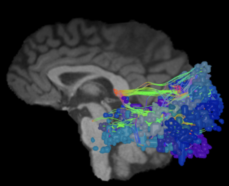
ᐅ SummaryArea FFC (fusiform face complex): part of basal occipital areas. Information hub receiving extensive input from the ventral stream about the static and dynamic details of invariant facial features and integrates them to produce the holistic form of a face. Plays an integral role in identity recognition and plays a minor role in emotional expression recognition due to its high activity for both neutral and expressive faces. ᐅ Where is it?Area FFC (fusiform face complex) is found in the posterior fusiform gyrus. It comprises the lateral half of the posterior portion of the gyrus and forms the medial bank of the adjacent portion of the occipitotemporal sulcus. ᐅ What are its borders?Area FFC borders TF anteriorly, TE2p and PH laterally, and V8 and PIT posteriorly. VVC forms its medial boundary. ᐅ What are its functional connections?Area FFC demonstrates functional connectivity to areas 1,2, 3a, and 3b in the sensory strip, area 4 in the motor strip, areas SCEF, FEF and 6v in the premotor region, areas OP4, STV, PFcm, 43, RI, LBelt, PBelt, A4, and A5 in the insula and opercular region, area TE2p in the temporal lobe, areas 7PC, PGp, PFt, AIP, MIP, VIP, LIPv, DVT, IP0, and IPS1 in the parietal lobe, areas V1, V2, V3, and V4 in the medial occipital lobe, areas V3a, V3b, V7, V6, and V6a of the dorsal visual stream, areas VVC, V8, PIT, VMV1, VMV2, and VMV3 of the ventral visual stream, and areas TPOJ1, TPOJ2, TPOJ3, V3cd, V4t, MT, LO1, LO2, LO3, MT, MST, PH, and FST of the lateral occipital lobe. ᐅ What are its white matter connections?Area FFC is structurally connected to the ILF. It also has many local U-fibers projecting from the parcellation. In some brains, fiber bundles connect with the SLF but this tract is inconsistent. The ILF projects toward the temporal pole and terminates at TGv. Short association bundles are connected to V4, PIT, LO3, FST, MST, MT, PH, and TPOJ2. ᐅ What is known about its function?Area FFC is an information hub receiving extensive input from the ventral stream about the static and dynamic details of invariant facial features and integrates them to produce the holistic form of a face. Using this information, area FFC plays an integral role in identity recognition and plays a minor role in emotional expression recognition due to its high activity for both neutral and expressive faces. |
|
A: lateral-medial
B: anterior-posterior
C: superior-inferior
DTI image |


ᐅ SummaryArea FST: part of the lateral surface areas of the occipital lobe. Acts as a major hub in the visual system, integrating massive amounts of visual information from both the dorsal and ventral streams. Plays an integral role in the perception of image content by integrating detail, motion and form-sensitive information; processing of spatial reference frames leading to continuous global-motion perception and spatial map formation; and stimulus filtering due to heightened attention-based motion selectivity. ᐅ Where is it?Area FST is found is an oblique to slightly vertically oriented area in the anterior portion of the lateral occipital lobe. It is just posterior to the end of the middle temporal gyrus. ᐅ What are its borders?Area FST borders MST and V4t posteriorly, and PHT anteriorly. PH is its inferior border and TPOJ2 is its superior border. ᐅ What are its functional connections?Area FST demonstrates functional connectivity to areas 1,2,3a, and 3b in the sensory strip, area 4 in the motor strip, areas SCEF, FEF, PEF, 6mp, 6r, 6v, 6a,and 6d in the premotor region, areas 24dd, p32prime, 23c, and 5mv in the cingulate regions, areas FOP2, OP1, OP4, PFcm, PoI1, PoI2, 43, RI, LBelt, PBelt, and A4 in the insula and opercular region, areas TE2p and PHT, in the temporal lobe, areas 7PC, 7AL, 7PL, 7am, PFt, PFop, PFm, AIP, VIP, LIPv, DVT, IP0, and IPS1 in the parietal lobe, areas V2, V3, and V4 in the medial occipital lobe, areas V3a, V3b, V7, V6, and V6a of the dorsal visual stream, areas FFC, VVC, V8, PIT, VMV1, VMV2, VMV3 of the ventral visual stream, and areas TPOJ2, TPOJ3, MT, MST, V3cd, V4t, MT, LO1, LO2, LO3, and PH of the lateral occipital lobe. ᐅ What are its white matter connections?Area FST is structurally connected with the SLF and ILF. SLF projections are consistent and terminate at the premotor area at parcellation 8C. ILF projections are inconsistent. Short association bundles are connected to PHT, LO1, LO2, LO3, MST, MT and PH, V4 ᐅ What is known about its function?Area FST acts as a major hub in the visual system, integrating massive amounts of visual information from both the dorsal and ventral streams. Area FST plays an integral role in the perception of image content by integrating detail, motion and form-sensitive information; processing of spatial reference frames leading to continuous global-motion perception and spatial map formation; and stimulus filtering due to heightened attention-based motion selectivity. |
|
A: lateral-medial
B: anterior-posterior
C: superior-inferior
DTI image |


ᐅ SummaryIPS1 (intraparietal sulcus 1): part of the lateral parietal lobe regions. Involved in processing object-based information. This suggests that it is important in object constancy, which describes the ability of an individual to interpret object permanence. Important in mentally visualizing the process of grasping objects as well as action associations and object function. Also plays a role in the transmission of top-down spatial attention information to the visual cortex during sustained attention tasks. ᐅ Where is it?Area IPS1 (Intraparietal sulcus 1) is found on the posterior, superior bank of the intraparietal sulcus. It should not be confused with IP1, which is on the inferior bank of the sulcus. IPS1 is the posterior most region on the superior bank of the intraparietal sulcus. ᐅ What are its borders?Area IPS1 borders IP0 inferiorly, MIP anteriorly, DVT medially, and area 7PL anteromedially. Its posterior border is made up of the visual areas V6a, V7 and V3b, which are part of the dorsal visual stream. ᐅ What are its functional connections?Area IPS1 demonstrates functional connectivity to areas 1 and 2 in the sensory strip, area 4 in the motor strip, areas SCEF and FEF in the premotor regions, areas TE2p and PHT in the temporal lobe, areas AIP, MIP, VIP, LIPd, LIPv, PGp, PFt, IP0, 7PL, and 7PC, in the lateral parietal lobe, areas DVT in the medial parietal lobe, areas V1, V2, V3, and V4 in the medial occipital lobe, areas V3a, V3b, V7, V6, and V6a of the dorsal visual stream, areas V8, PIT, FFC, VVC, VMV1, VMV2, and VMV3 of the ventral visual stream, and areas V3cd, V4t, LO1, LO2, LO3, PH, TPOJ2, MT, MST and FST of the lateral occipital lobe ᐅ What are its white matter connections?Area IPS1 is structurally connected to the inferior fronto- occipital fasciculus (IFOF), MdLF and local parcellations. The IFOF courses from IPS1 through the posterior temporal lobe and extreme/external capsule to frontal lobe parcellations. IFOF terminations are inconsistent across individuals but the majority end at the superior temporal gyrus involving parcellations 8BL, 9a and 9p. Connections with the MdLF travel deep to the parietal lobe to the superior temporal gyrus and planum temporale to end at A4, PBelt and MBelt. Local short association fibers connect with MIP, V7, V6A and V3B. ᐅ What is known about its function?Area IPS1 is involved in processing object-based information. This suggests that this region is important in object constancy, which describes the ability of an individual to interpret object permanence. This region is important in mentally visualizing the process of grasping objects as well as action associations and object function. Area IPS1 also plays a role in the transmission of top-down spatial attention information to the visual cortex during sustained attention tasks. |
|
A: lateral-medial
B: anterior-posterior
C: superior-inferior
DTI image |


ᐅ SummaryArea LO1 (lateral occipital 1): part of the lateral surface areas of the occipital lobe. Higher order visual structure that receives detail, motion, and characteristic information about the central visual field from both the dorsal and ventral streams. Preferentially activates in response to orientation-selective and boundary information to process the form of objects. ᐅ Where is it?Area LO1 (Lateral occipital 1) is a small vertically oriented area in the posterior occipital lobe, just anterior to the occipital pole. ᐅ What are its borders?Area LO1 borders V4 posteriorly, and LO3 anteriorly. Its superior border is made up of V3cd. V4t and LO2 form its inferior border ᐅ What are its functional connections?Area LO1 demonstrates functional connectivity to area FEF in the premotor region, area A4 in the insula and opercular region, areas VIP, LIPv, IP0, DVT, and IPS1 in the parietal lobe, areas V1, V2, V3, and V4 in the medial occipital lobe, areas V3a, V3b, V7, V6, and V6a of the dorsal visual stream, areas FFC, VVC, V8, PIT, VMV1, VMV2, VMV3 of the ventral visual stream, and areas V3cd, V4t, MT, LO2, LO3, PH, and FST of the lateral occipital lobe. ᐅ What are its white matter connections?Area LO1 is structurally connected to local parcellations. Short association bundles are connected to V3A, V3B, V3CD, LO2, and LO3 ᐅ What is known about its function?Area LO1 is a higher-order visual structure that receives detail, motion, and characteristic information about the central visual field from both the dorsal and ventral streams. Area LO1 preferentially activates in response to orientation-selective and boundary information to process the form of objects. |
|
A: lateral-medial
B: anterior-posterior
C: superior-inferior
DTI image |


ᐅ SummaryArea LO2 (lateral occipital 2): part of the lateral surface areas of the occipital lobe. hows preferential retinotopic activation in the peripheral visual field, and integrates inputs from the ventral and dorsal streams to encode information about the shapes of stimuli. This region also demonstrates high activation in the processing, encoding, and recognition of objects. ᐅ Where is it?Area LO2 (Lateral occipital 2) is a small horizontally oriented area just anterior to the occipital pole and slightly superior to the tentorium. ᐅ What are its borders?Area LO2 borders V4 posteriorly, and PH anteriorly. Its superior border is V4t and its inferior border is PIT. ᐅ What are its functional connections?Area LO2 demonstrates functional connectivity to area FEF in the premotor region, area PBelt in the insula and opercular region, areas VIP, LIPv, and IPS1 in the parietal lobe, areas V1, V2, V3, and V4 in the medial occipital lobe, areas V3a, V3b, V7, V6, and V6a of the dorsal visual stream, areas FFC, VVC, V8, PIT, VMV3 of the ventral visual stream, and areas V3cd, V4t, MT, LO1, LO3, PH, and FST of the lateral occipital lobe. ᐅ What are its white matter connections?Area LO2 is structurally connected to local parcellations. Short association bundles are connected to V3A, V3B, V3CD, LO1, and LO3 ᐅ What is known about its function?Area LO2 shows preferential retinotopic activation in the peripheral visual field, and integrates inputs from the ventral and dorsal streams to encode information about the shapes of stimuli. This region also demonstrates high activation in the processing, encoding, and recognition of objects. |
|
A: lateral-medial
B: anterior-posterior
C: superior-inferior
DTI image |


ᐅ SummaryArea LO3 (lateral occipital 3): part of the lateral surface areas of the occipital lobe. Literature suggests this region of the occipital lobe is an important hub between the dorsal and ventral streams to integrate, encode, and process detail, motion, and shape information for object recognition and encoding. ᐅ Where is it?Area LO3 (lateral occipital 3) is a vertically oriented area in the superior central portion of the lateral occipital lobe. It is located just inferior to the posterior angular gyrus. ᐅ What are its borders?Area LO3 borders V3cd and LO1 posteriorly, and MT anteriorly. Its superior border is made up of TPOJ3 and PGp. Its inferior border is made up of LO1 and V4t. ᐅ What are its functional connections?Area LO3 demonstrates functional connectivity to areas 1,2, 3a, and 3b in the sensory strip, area 4 in the motor strip, areas SCEF and FEF in the premotor region, areas 5mv and 23c in the cingulate regions, areas OP4, PFcm, 43, RI, PBelt, and A4 in the insula and opercular region, areas 7PC, DVT, PFop, PGp, VIP, LIPv, IP0, and IPS1 in the parietal lobe, areas V1, V2, V3, and V4 in the medial occipital lobe, areas V3a, V3b, V7, V6, and V6a of the dorsal visual stream, areas FFC, VVC, V8, PIT, VMV1, VMV2, and VMV3 of the ventral visual stream, and areas TPOJ2, TPOJ3 V3cd, V4t, MT, LO1, LO2, PH, and FST of the lateral occipital lobe. ᐅ What are its white matter connections?Area LO3 is structurally connected to the ILF and local parcellations. ILF projections travel through the temporal lobe to terminate at TGv. Short association bundles are connected to V3A, V3B, V3CD, LO1, LO2, PH and FST. ᐅ What is known about its function?While LO3 is considered a new area, the literature suggests this region of the occipital lobe is an important hub between the dorsal and ventral streams to integrate, encode, and process detail, motion, and shape information for object recognition and encoding. |
|
A: lateral-medial
B: anterior-posterior
C: superior-inferior
DTI image |


ᐅ SummaryArea MST (medial superior temporal) part of the lateral surface areas of the occipital lobe. Receives direct, functional input from area MT and is responsible for the integration and analysis of global, visual motion and the perception of self-motion. Involved in the execution and continuation of smooth pursuit eye movements, in coordination with the frontal eye fields. ᐅ Where is it?Area MST (medial superior temporal area) is a vertically oriented area found paralleling and just anterior to MT ᐅ What are its borders?just below the angular gyrus. ᐅ What are its functional connections?Area MST borders MT posteriorly ᐅ What are its white matter connections?and FST anteriorly. Its superior border is made up of TPOJ2 and TPOJ3. Its inferior border is formed by FST and V4t. ᐅ What is known about its function?Area MST demonstrates functional connectivity to areas 1 |
|
A: lateral-medial
B: anterior-posterior
C: superior-inferior
DTI image |


ᐅ SummaryArea MT (middle temporal): part of the lateral surface areas of the occipital lobe. Neurons respond to direction-sensitive visual motion and are responsible for the integration of one-dimensional visual signals into a two- dimensional visual motion pattern, binocular disparity tuning, noise reduction, segmentation of figure and background in complex and moving stimuli, and initiation of smooth-pursuit eye movements. ᐅ Where is it?Area MT (Middle temporal area) is a vertically oriented area in the superior part of the central lateral occipital lobe. It is located just inferior to the angular gyrus. ᐅ What are its borders?Area MT borders LO3 posteriorly, and MST anteriorly. Its superior border is formed by TPOJ3 and its inferior border by V4t. ᐅ What are its functional connections?Area MT demonstrates functional connectivity to areas 1,2,3a, and 3b in the sensory strip, area 4 in the motor strip, areas OP4, RI, PBelt, A4, and A5 in the insula and opercular region, areas VIP, LIPv, and IPS1 in the parietal lobe, areas V2, V3, and V4 in the medial occipital lobe, areas V3a, V3b, V7, V6, and V6a of the dorsal visual stream, areas FFC, VVC, V8, PIT, VMV3 of the ventral visual stream, and areas V3cd, V4t, MST, LO1, LO2, LO3, PH, and FST of the lateral occipital lobe. ᐅ What are its white matter connections?Area MT is structurally connected with the ILF. These projections are inconsistent across brains. ILF projections travel through the temporal lobe to end at TF. There are many short association bundles connecting to MST, LO1, LO2, LO3, TPOJ2, TPOJ3, FST, PH, V3b and IPO. ᐅ What is known about its function?Neurons in area MT respond to direction-sensitive visual motion and are responsible for the integration of one-dimensional visual signals into a two- dimensional visual motion pattern, binocular disparity tuning, noise reduction, segmentation of figure and background in complex and moving stimuli, and initiation of smooth-pursuit eye movements. |
|
A: lateral-medial
B: anterior-posterior
C: superior-inferior
DTI image |


ᐅ SummaryArea PIT (posterior infratemporal cortex): part of basal occipital areas. Implicated in the analysis of the basic characteristics of color, such as hue, saturation, and brightness based on information provided by V8. ᐅ Where is it?Area PIT (Posterior infratemporal cortex) is found posterior most portion of the occipitotemporal gyrus where it begins to blend into the occipital pole. It spills slightly onto the lateral surface. ᐅ What are its borders?Area PIT borders V8 and V4 medially. V4 also forms its posterior boundary. It borders FFC and PH anteriorly. LO2 is its superior (lateral) neighbor. ᐅ What are its functional connections?Area PIT demonstrates functional connectivity to area FEF in the premotor region, areas PBelt, and A4 in the insula and opercular region, areas VIP, LIPv, IPS1 and DVT in the parietal lobe, areas V1, V2, V3, and V4 in the medial occipital lobe, areas V3a, V3b, V7, V6, and V6a of the dorsal visual stream, areas VVC, FFC, V8 of the ventral visual stream, and areas V3cd, V4t, LO1, LO2, LO3, PH, MST, and FST of the lateral occipital lobe. ᐅ What are its white matter connections?Area PIT is structurally connected to VOF and its surrounding parcellations. VOF connections project mediodorsally to terminate at V3, V2 and V3a. Short association bundles are connected to PH, V8, V4 and V1. ᐅ What is known about its function?Area PIT has been implicated in the analysis of the basic characteristics of color, such as hue, saturation, and brightness based on information provided by V8. |
|
A: lateral-medial
B: anterior-posterior
C: superior-inferior
DTI image |


ᐅ SummaryArea PIT (posterior infratemporal cortex): part of basal occipital areas. Implicated in the analysis of the basic characteristics of color, such as hue, saturation, and brightness based on information provided by V8. ᐅ Where is it?Area PIT (Posterior infratemporal cortex) is found posterior most portion of the occipitotemporal gyrus where it begins to blend into the occipital pole. It spills slightly onto the lateral surface. ᐅ What are its borders?Area PIT borders V8 and V4 medially. V4 also forms its posterior boundary. It borders FFC and PH anteriorly. LO2 is its superior (lateral) neighbor. ᐅ What are its borders?Area PIT demonstrates functional connectivity to area FEF in the premotor region, areas PBelt, and A4 in the insula and opercular region, areas VIP, LIPv, IPS1 and DVT in the parietal lobe, areas V1, V2, V3, and V4 in the medial occipital lobe, areas V3a, V3b, V7, V6, and V6a of the dorsal visual stream, areas VVC, FFC, V8 of the ventral visual stream, and areas V3cd, V4t, LO1, LO2, LO3, PH, MST, and FST of the lateral occipital lobe. ᐅ What are its functional connections?Area PIT is structurally connected to VOF and its surrounding parcellations. VOF connections project mediodorsally to terminate at V3, V2 and V3a. Short association bundles are connected to PH, V8, V4 and V1. ᐅ What are its white matter connections?Area PIT has been implicated in the analysis of the basic characteristics of color, such as hue, saturation, and brightness based on information provided by V8. |
|
A: lateral-medial
B: anterior-posterior
C: superior-inferior
DTI image |
ᐅ SummaryArea V1 (visual area 1): part of occipital lobe. Primary visual cortex. Heavily implicated in the initial detection of motion, and signaling to the middle temporal area for the directional and spatial integration of global motion. ᐅ Where is it?Area V1 (Visual area 1) is the primary visual cortex, which is located on the banks of the calcarine sulcus. It fills both banks of the sulcus and extends onto the tip of the occipital pole. ᐅ What are its borders?Area V1 borders V2 along its entire length. Its anterior border is with the prostriate cortex. ᐅ What are its functional connections?Area V1 demonstrates functional connectivity to areas SCEF and FEF in the premotor region, areas 46, 9-46d, p9-46v, 23c, 5mv, a24prime, p32prime, a32prime, and p24 in the frontal lobe, areas FOP4, FOP5, AVI, PoI1, 43, LBelt, PBelt, and A1 in the insula and opercular region, area PHT in the temporal lobe, areas 7am, 7PL, PGp, PFop, PF, MIP, LIPd, LIPv, IPS1, IP1, IP0, POS2, RSC, PCV, and DVT in the parietal lobe, areas ProS, V2, V3, and V4 in the medial occipital lobe, areas V3a, V3b, V7, V6, and V6a of the dorsal visual stream, areas FFC, VVC, V8, PIT, VMV1, VMV2 and VMV3 of the ventral visual stream, and areas V3cd, LO1, LO2, LO3, PH, and FST of the lateral occipital lobe (Figure 3). ᐅ What are its white matter connections?Area V1 is structurally connected to the IFOF, MdLF, optic radiations (OR) and splenium of the corpus callosum. IFOF projections terminate at parcellations in the frontal pole including 11I, a10p and p10p. Connections from the MdLF terminate in the superior temporal gyrus at STSda. There are consistent connections with FM to contralateral V1 through the splenium of the corpus callosum. Short association bundles are connected to V2, V3, and V3B (Figure ᐅ What is known about its function?In each hemisphere, Area V1 encodes an inverted, contralateral hemifield with greater cortical surface area devoted to visual stimuli detected by the fovea, indicating greater processing of input closer to the center of the visual field. It has also been heavily implicated in the initial detection of motion, and signaling to the Middle Temporal Area (MT) for the directional and spatial integration of global motion. |
|
A: lateral-medial
B: anterior-posterior
C: superior-inferior
DTI image |


ᐅ SummaryArea V2 (visual area 2): part of occipital lobe. Implicated in more complex visual integration after receiving information from V1, most notably, the differentiation between stimulus and background. ᐅ Where is it?Area V2 (Visual area 2) is "C" shaped and wraps around V1. Its apex loops slightly around the occipital pole onto the lateral surface. Its upper limb is slightly shorter but makes up a large fraction of the cuneus. Its inferior limb makes up a large part of the lingula. ᐅ What are its borders?Area V2 wraps around V1 interiorly, and V3 makes up its external boundary. V6 and DVT make up the anterior border of its upper limb, and the anterior tip of its inferior limb forms a wedge between ProS and VMV1. ᐅ What are its functional connections?Area V2 demonstrates functional connectivity to areas 1, 2, 3a, 3b in the sensory strip, area 4 in the motor strip, areas SCEF, FEF and 6a in the premotor region, areas 46, 9-46d, 23c, 5mv, 24dd, 24dv, a24prime, and p32prime in the frontal lobe, areas FOP3, FOP4, OP4, OP2-3, PoI1, PoI2, PFcm, 43, 52, RI, TA2, STV, PSL, MBelt, LBelt, PBelt, A1, and A4 in the insula and opercular region, area PHT in the temporal lobe, areas 7PC, PGp, PFop, AIP, VIP, MIP, LIPd, LIPv, IPS1, IP0, PCV, and DVT in the parietal lobe, areas ProS, V1, V3, and V4 in the medial occipital lobe, areas V3a, V3b, V7, V6, and V6a of the dorsal visual stream, areas FFC, VVC, V8, PIT, VMV1, VMV2 and VMV3 of the ventral visual stream, and areas TPOJ1, TPOJ2, TPOJ3, MST, MT, V3cd, V4t, LO1, LO2, LO3, PH, and FST of the lateral occipital lobe. ᐅ What are its white matter connections?Area V2 is structurally connected to the IFOF, ILF, MdLF and contralateral hemisphere. IFOF projections terminate at parcellations in the frontal pole including 8BL, 9a, 9p, 10d, 10pp, a10p and 10pp. Connections from the MdLF terminate in the superior temporal gyrus at STSda, TGd, and TE1a. There are consistent connections with the FM to the contralateral hemisphere through the splenium of the corpus callosum that connect to V1, V2, V3, V4, V6, DVT and V3A. Short association bundles are connected to V1, V2, V3, V4, V6, V3A and DVT. ᐅ What is known about its function?Area V2 partitions the visual field into two discontinuous, inverted, contralateral quarterfield maps divided by the horizontal meridian. V2 also perceives visual stimuli at greater angles from the foveal point than V1, indicating its perception of the visual field is farther from the fovea than V1. Additionally, V2 has been implicated in more complex visual integration after receiving information from V1, most notably, the differentiation between stimulus and background. |
|
A: lateral-medial
B: anterior-posterior
C: superior-inferior
DTI image |


ᐅ SummaryArea V3 (visual area 3): part of occipital lobe. Shown to play a role in the perception and integration of global motion by processing directionally specific, visual, local motion in its broad receptive fields and transmitting information to area V6, and the dorsal visual stream for further processing. ᐅ Where is it?Area V3 (Visual area 3) is "C" shaped and wraps around V2. Its apex loops around the occipital pole well onto the lateral surface. Its upper limb makes up a lesser fraction of the cuneus. Its inferior limb makes up the lower half of the lingula. ᐅ What are its borders?Area V3 wraps around V2 interiorly, and V4 makes up its external boundary. Its anterior limb has V3a as a superior boundary and V6 as an anterior boundary. Its inferior limb has VMV1 as an anterior boundary. ᐅ What are its functional connections?Area V3 demonstrates functional connectivity to areas 1, 2, 3a, 3b in the sensory strip, area 4 in the motor strip, areas SCEF, FEF and 6a in the premotor region, areas 46, 9-46d, 23c, 5mv, a24prime, and p32prime in the frontal lobe, areas FOP3, FOP4, OP4, OP2-3, PoI1, PFcm, 43, 52, RI, TA2, STV, PSL, MBelt, LBelt, PBelt, A1, and A4 in the insula and opercular region, areas PGp, PFop, VIP, LIPd, LIPv, IPS1, IP0, and DVT in the parietal lobe, areas ProS, V1, V3, and V4 in the medial occipital lobe, areas V3a, V3b, V7, V6, and V6a of the dorsal visual stream, areas FFC, VVC, V8, PIT, VMV1, VMV2 and VMV3 of the ventral visual stream, and areas TPOJ1, TPOJ2, ,MST, MT, V3cd, V4t, LO1, LO2, LO3, PH, and FST of the lateral occipital lobe. ᐅ What are its white matter connections?Area V3 is structurally connected to the IFOF, ILF, MdLF and FM. The IFOF projections originate from the superior portion of the parcellation before it curves inferiorly to the basal surface of the occipital lobe. Projections from the IFOF have typical anterior terminations that include 8BL, 9a, 9p and 10s. Connections from the MdLF terminate at A5 of the superior temporal gyrus. ILF projections originate from the inferior portion of V3 along the basal surface of the occipital lobe and terminate at STGa and TGd. Contralateral connections with FM course through the splenium of the corpus callosum and terminate at V1 and V2. Short association bundles are connected to V1, V2, V3A and V4 ᐅ What is known about its function?Similar to V2, Area V3 receives visual input as two discontinuous, inverted, contralateral quarterfield maps divided by the horizontal meridian. Area V3 perceives even wider degrees of the visual field than V2, indicating it perceives visual stimuli at an even further angle from the fovea than V2. Additionally, Area V3 (and in a more limited role, V2) has been shown to play a role in the perception and integration of global motion by processing directionally specific, visual, local motion in its broad receptive fields and transmitting information to Area V6 and the dorsal visual stream for further processing. |
|
A: lateral-medial
B: anterior-posterior
C: superior-inferior
DTI image |


ᐅ SummaryArea V3a (visual area 3a): part of the superior surface areas of the occipital lobe. Demonstrates high motion sensitivity and high contrast sensitivity in the central visual field surrounding the foveal point indicating its role in motion-selective visual perception. This region also integrates spatial information from contralateral and ipsilateral visual input sources. ᐅ Where is it?Area V3a (Visual area 3a) is found in the superior surface of the occipital lobe, near the interhemispheric fissure. It is positioned similar to the superior limb of V4, which as described above, is hypoplastic compared to V3. ᐅ What are its borders?Area V3a borders V3 inferiorly, and V4 posteriorly. Its lateral border is V3b. Its anterior border included V7 and V6a. V6 is its anterior neighbor across the interhemispheric cleft. ᐅ What are its functional connections?Area V3a demonstrates functional connectivity to areas 1, 3a, 3b in the sensory strip, area 4 in the motor strip, areas SCEF, FEF in the premotor region, areas a24prime, and p32prime, 5mv, 23c in the medial frontal lobe, areas FOP1, FOP3, FOP4, OP4, OP2-3, 43, PFcm, PoI1, RI, 52, A4, LBelt, and PBelt in the insula opercular regions, areas VIP, LIPv, PGp, PFop, IPS1, IP0, and DVT in the parietal lobe, areas ProS, V1, V2, V3, and V4 in the medial occipital lobe, areas V3b, V7, V6, and V6a of the dorsal visual stream, areas FFC, VVC, V8, VMV1, VMV2, and VMV3 of the ventral visual stream, and areas TPOJ2, V3cd, V4t, LO1, LO2, LO3, MT, MST, PH, and FST of the lateral occipital lobe. ᐅ What are its white matter connections?Area V3a is structurally connected to the IFOF, MdLF and FM. The IFOF has consistent terminations to the frontal lobe at parcellations 8BL, 9a, 9p, 9m, 10pp and 10d. There are projections from the MdLF to the superior temporal gyrus that terminate at STGa. FM connections course through the splenium of the corpus callosum to terminate at contralateral V3a. V3a has short association bundles connected to V6A, V3b, V6, V7, and V3. Right hemisphere has inconsistent projections with the IFOF when compared to left hemisphere. ᐅ What is known about its function?Area V3a demonstrates high motion sensitivity and high contrast sensitivity in the central visual field surrounding the foveal point indicating its role in motion-selective visual perception. This region also integrates spatial information from contralateral and ipsilateral visual input sources. |
|
A: lateral-medial
B: anterior-posterior
C: superior-inferior
DTI image |


ᐅ SummaryArea V3b (visual area 3b): part of the superior surface areas of the occipital lobe. Acts as a relay point, collecting motion-sensitive information from the dorsal stream, and is implicated in the processing of motion information and discerning the edges of moving objects, known as kinetic boundaries. ᐅ Where is it?Area V3b (Visual area 3b) is found on the superior occipital surface. ᐅ What are its borders?Area V3b borders IP0 anterolaterally, IPS1 anteromedially, and V7 medially. Its posterior border is made up of V3a and the superior limb of V4. V3cd is its lateral neighbor. ᐅ What are its functional connections?Area V3b is connected to areas SCEF, FEF in the premotor region, areas a24prime, and p32prime, 23c in the medial frontal lobe, areas 43, RI, and PBelt in the insula opercular regions, areas 7PC, VIP, IPS1, IP0, and DVT in the parietal lobe, areas ProS, V1, V2, V3, and V4 in the medial occipital lobe, areas V3a, V7, V6, and V6a of the dorsal visual stream, areas FFC, VVC, V8, VMV1, VMV2, and VMV3 of the ventral visual stream, and areas V3cd, V4t, LO1, LO2, LO3, MT, MST, PH, and FST of the lateral occipital lobe. ᐅ What are its white matter connections?Area V3b is structurally connected with local parcellations. There are connections with IFOF and MdLF but this is inconsistent across brains. Short association bundles are connected to MIP, IPO, IPS1, V4 and V3CD. ᐅ What is known about its function?Area V3b acts as a relay point, collecting motion-sensitive information from the dorsal stream, and is implicated in the processing of motion information and discerning the edges of moving objects, known as kinetic boundaries. |
|
A: lateral-medial
B: anterior-posterior
C: superior-inferior
DTI image |


ᐅ SummaryAreaV3cd (visual area 3 c/d): part of the lateral surface areas of the occipital lobe. Integrates object detail information, such as contrast and kinetic edges, to create an holistic image for object recognition. ᐅ Where is it?Area V3cd (Visual area 3 c/d) is a vertical strip in the posterosuperior portion of the lateral occipital lobe, just posterior to the angular gyrus. ᐅ What are its borders?Area V3cd borders PGp and LO3 anteriorly, LO1 inferiorly, V4 posteriorly, and V3b superiorly. ᐅ What are its functional connections?Area V3cd demonstrates functional connectivity to area FEF in the premotor region, area PBelt in the insula and opercular region, areas 7PC, AIP, VIP, LIPv, PGp, IPS1, IP0, and DVT in the parietal lobe, areas ProS, V1, V2, V3, and V4 in the medial occipital lobe, areas V3a, V3b, V7, V6, and V6a of the dorsal visual stream, areas VVC, V8, VMV1, VMV2, and VMV3 of the ventral visual stream, and areas V3cd, MT, MST, V4t, LO1, LO2, LO3, PH, and FST of the lateral occipital lobe. ᐅ What are its white matter connections?Area V3cd is structurally connected to local parcellations. Short association bundles are connected to V3B, V7, V4t, V3CD, LO1, LO2, LO3, MST, and MT). ᐅ What is known about its function?Area V3cd integrates object detail information, such as contrast and kinetic edges, to create an holistic image for object recognition. |
|
A: lateral-medial
B: anterior-posterior
C: superior-inferior
DTI image |


ᐅ SummaryArea V4 (visual area 4): part of occipital lobe. Shown to encode texture, patterns, form, and color perception, making it integral to object and pattern recognition. Literature has also implicated V4 in the perception of contralateral, hemifield representations of the peripheral visual field. ᐅ Where is it?Area V4 (Visual area 4) is "C" shaped and wraps around V3. It is primarily located on the posterior aspect of the lateral occipital surface. Its upper limb does not extend onto the medial surface as it's somewhat shorter than the corresponding limbs of V3 and V2. Its inferior limb extends onto the medial basal surface of the occipital lobe. ᐅ What are its borders?Area V4 borders V3 internally. Its superior limb ends in V3a and its inferior limb ends in VMV2 and VMV3. It shares an external border with a large number of visual processing areas. From superior to inferior these are: V3b, V3cd, LO1, LO2, Pit and V8. ᐅ What are its functional connections?Area V4 demonstrates functional connectivity to areas 1, 3a, 3b in the sensory strip, area 4 in the motor strip, areas SCEF, FEF and 6a in the premotor region, areas 9-46d, 23c, a24prime, and p32prime in the frontal lobe, areas FOP3, OP4, PFcm, 43, 52, RI, TA2, STV, PSL, MBelt, LBelt, PBelt, A1, and A4 in the insula and opercular region, areas PFop, VIP, LIPv, IPS1, IP0, and DVT in the parietal lobe, areas ProS, V1, V3, and V4 in the medial occipital lobe, areas V3a, V3b, V7, V6, and V6a of the dorsal visual stream, areas FFC, VVC, V8, PIT, VMV1, VMV2 and VMV3 of the ventral visual stream, and areas TPOJ1, MST, MT, V3cd, V4t, LO1, LO2, LO3, PH, and FST of the lateral occipital lobe. ᐅ What are its white matter connections?Area V4 is structurally connected with the ILF, MdLF and portions of the IFOF. ILF projections terminate at TF. IFOF projections are inconsistent and the parcellations at which this tract terminates are unable to be discerned. MdLF connections terminate at the superior temporal gyrus at A4 and A5. Short association bundles are connected to V3, V3A, V3B, V3CD, LO1, LO2, and V6A. ᐅ What is known about its function?Area V4 has been shown to encode texture, patterns, form, and color perception, making it integral to object and pattern recognition. Interestingly, the literature has also implicated V4 in the perception of contralateral, hemifield representations of the peripheral visual field. |
|
A: lateral-medial
B: anterior-posterior
C: superior-inferior
DTI image |


ᐅ SummaryArea V4t (visual area 4t): part of the lateral surface areas of the occipital lobe. Shown in the literature to integrate information from both the ventral and dorsal streams, and demonstrates a high level of activity in response to both motion and shape-sensitive information, indicating its significance in the integration of object processing and global-motion perception. ᐅ Where is it?Area V4t (Visual area 4t) is a horizontal area in the central portion of the lateral occipital cortex. ᐅ What are its borders?Area V4t borders LO1 posteriorly, and FST anteriorly. Its inferior border is LO2. Its superior border is made up of MT, MST, and LO3. ᐅ What are its functional connections?Area V4t demonstrates functional connectivity to areas 1,2,3a, and 3b in the sensory strip, area 4 in the motor strip, area FEF in the premotor region, areas RI, PBelt, and A4 in the insula and opercular region, areas VIP, LIPv, and IPS1 in the parietal lobe, areas V2, V3, and V4 in the medial occipital lobe, areas V3a, V3b, V7, V6, and V6a of the dorsal visual stream, areas VVC, V8, VMV1, VMV2, and VMV3 of the ventral visual stream, and areas V3cd, MT, MST, LO3, PH, and FST of the lateral occipital lobe. ᐅ What are its white matter connections?Area V4t is structurally connected with local parcellations. Tracts originating from this region such as SLF and ILF are inconsistent across brains. Short association bundles are connected to V4t, LO3, MST, MT, LO1 and V3CD. ᐅ What is known about its function?Area V4t has been shown in the literature to integrate information from both the ventral and dorsal streams, and demonstrates a high level of activity in response to both motion and shape-sensitive information, indicating its significance in the integration of object processing and global-motion perception. |
|
A: lateral-medial
B: anterior-posterior
C: superior-inferior
DTI image |


ᐅ SummaryArea V6 (visual area 6): part of the superior surface areas of the occipital lobe. Implicated in the processing and analysis of visual motion and has been demonstrated in lesion studies to cause motion blindness and other motion-related visual disturbances. ᐅ Where is it?Area V6 (Visual area 6) is a vertically oriented area in the anterosuperior portion of the cuneus, just posterior to the superior parieto-occipital sulcus. ᐅ What are its borders?Area V6 borders the superior limbs of V2 and V3 posteriorly, and DVT anteriorly. V2 is its inferior boundary, and V6a is its superior boundary. ᐅ What are its functional connections?Area V6 demonstrates functional connectivity to areas 1, 2, 3a, 3b in the sensory strip, area 4 in the motor strip, areas SCEF, FEF in the premotor region, areas 9-46d, and 46 in the lateral frontal lobe, areas a24prime, and p32prime, 5mv, 23c in the medial frontal lobe, areas FOP1, FOP3, FOP4, OP4, OP2-3, 43, PFcm, STV, PoI1, PoI2, MI, RI, TA2, 52, A4, MBelt, and PBelt in the insula opercular regions, areas 7PC, 7AL, 7am, VIP, LIPv, PGp, PFop, IPS1, IP0, PCV, and DVT in the parietal lobe, areas ProS, V1, V2, V3, and V4 in the medial occipital lobe, areas V3a, V3b, V7, and V6a of the dorsal visual stream, areas FFC, VVC, V8, VMV1, VMV2, and VMV3 of the ventral visual stream, and areas TPOJ2, TPOJ3, V3cd, V4t, LO1, LO2, LO3, MT, MST, PH, and FST of the lateral occipital lobe. ᐅ What are its white matter connections?Area V6 is structurally connected to the IFOF, MdLF and FM. IFOF projections terminate at parcellelations in the frontal lobe including 9a, 9p, 9m and 8BL. The MdLF runs parallel to the IFOF then courses laterally to the superior temporal gyrus, as the IFOF courses medially between the lateral ventricle and insula. The MdLF projections terminate at MBelt, A5, and PI. FM connections travel through the splenium of the corpus callosum to terminate at V6, V2 and V1. Short association bundles are connected to V7, V3, V3b, and V3a. Right hemisphere has consistent ILF projections. ᐅ What is known about its function?Area V6 has been implicated in the processing and analysis of visual motion and has been demonstrated in lesion studies to cause motion blindness and other motion-related visual disturbances. |
|
A: lateral-medial
B: anterior-posterior
C: superior-inferior
DTI image |


ᐅ SummaryArea V6A (visual area 6a): part of the superior surface areas of the occipital lobe. Responds to peripheral visual stimuli and has been implicated in the integration and evaluation of spatial information to coordinate eye and arm movements, indicating its importance for reaching and hand¨Ceye coordination. ᐅ Where is it?Area V6A (Visual area 6a) is a small area which straddles the angle of the interhemispheric cleft of the superior occipital lobe, lying just posterior to the superior most limit of the parieto-occipital sulcus. ᐅ What are its borders?Area V6A borders V3a posteriorly, and DVT anteriorly. Its lateral border is made V7 and IPS1. V6 forms its inferior border. ᐅ What are its functional connections?Area V6A demonstrates functional connectivity to areas 1, 2, 3a, 3b in the sensory strip, area 4 in the motor strip, areas SCEF, FEF in the premotor region, areas a24prime, and p32prime, 5mv, 23c in the medial frontal lobe, areas OP4, 43, RI, A4, and PBelt in the insula opercular regions, areas 7PC, VIP, LIPv, PGp, IPS1, IP0, and DVT in the parietal lobe, areas ProS, V1, V2, V3, and V4 in the medial occipital lobe, areas V3a, V3b, V7, and V6 of the dorsal visual stream, areas FFC, VVC, V8, VMV1, VMV2, and VMV3 of the ventral visual stream, and areas TPOJ2, V3cd, V4t, LO1, LO2, LO3, MT, MST, PH, and FST of the lateral occipital lobe. ᐅ What are its white matter connections?Area V6A is structurally connected to the IFOF and MdLF. There are inconsistent projections with FM. IFOF projections terminate at frontal lobe parcellations 8BM and 8BL. The MdLF fibers run parallel to the IFOF then course laterally to the superior temporal gyrus to terminate at A4 and MBelt. Short association bundles are connected to V3B and V7. ᐅ What is known about its function?Area V6A responds to peripheral visual stimuli and has been implicated in the integration and evaluation of spatial information to coordinate eye and arm movements, indicating its importance for reaching and hand-eye coordination. |
|
A: lateral-medial
B: anterior-posterior
C: superior-inferior
DTI image |


ᐅ SummaryArea V7 (visual area 7): part of the superior surface areas of the occipital lobe. Contains a relatively narrow retinotopic map of the central visual field surrounding the foveal point and shows increased stimulation in response to attention directed toward a visual stimulus. Uses this visual information to integrate spatial information within the central visual field. ᐅ Where is it?Area V7 (Visual area 7) is found on the superior surface of the occipital lobe near the interhemispheric cleft, roughly lateral to the superior most point of the intraparietal sulcus. ᐅ What are its borders?Area V7 borders V3a posteriorly, and IPS1 anteriorly. Its lateral border is made up of V3b, and its medial border is formed by V6a. ᐅ What are its functional connections?Area V7 demonstrates functional connectivity to areas 1, 2, 3a, 3b in the sensory strip, area 4 in the motor strip, areas SCEF, FEF in the premotor region, areas a24prime, and p32prime, 23c in the medial frontal lobe, areas OP4, 43, MI, RI, A4, LBelt, and PBelt in the insula opercular regions, areas VIP, LIPv, IPS1, IP0, and DVT in the parietal lobe, areas ProS, V1, V2, V3, and V4 in the medial occipital lobe, areas V3a, V3b, V6, and V6a of the dorsal visual stream, areas FFC, VVC, V8, VMV1, VMV2, and VMV3 of the ventral visual stream, and areas V3cd, V4t, LO1, LO2, LO3, MT, MST, PH, and FST of the lateral occipital lobe. ᐅ What are its white matter connections?Area V7 is structurally connected with IFOF, MdLF and ILF. The termination of the IFOF is inconsistent across brains. The MdLF fibers run parallel to the IFOF then course laterally to the superior temporal gyrus to terminate at A5, STSda, STSdp, STSva and TGd. ILF projections travel through the temporal lobe to end at TGv and TGd. Short association bundles are connected to LO1, LO2, LO3, V3CD, V3A, V6A, and V6. ᐅ What is known about its function?Area V7 contains a relatively narrow retinotopic map of the central visual field surrounding the foveal point and shows increased stimulation in response to attention directed toward a visual stimulus. Area V7 uses this visual information to integrate spatial information within the central visual field. |
|
A: lateral-medial
B: anterior-posterior
C: superior-inferior
DTI image |


ᐅ SummaryArea V8 (visual area 8): part of basal occipital areas. Most notably responsible for the perception and processing of color in the visual field. ᐅ Where is it?Area V8 (Visual area 8) is found at the far posterior most portion of the fusiform gyrus. ᐅ What are its borders?Area V8 borders shares its entire medial and posterior border with V4. Its anteromedial border is with VMV3, its anterior border is with VVC, and anterolateral border is with FFC, which also forms some of its lateral border. PIT forms its posterolateral border. ᐅ What are its functional connections?Area V8 demonstrates functional connectivity to area FEF in the premotor region, areas 43, PBelt, and A4 in the insula and opercular region, areas VIP, LIPv, IPS1, IP0, and DVT in the parietal lobe, areas ProS, V1, V2, V3, and V4 in the medial occipital lobe, areas V3a, V3b, V7, V6, and V6a of the dorsal visual stream, areas VVC, V8, VMV2 and VMV3 of the ventral visual stream, and areas V3cd, LO1, LO3, PH, and FST of the lateral occipital lobe. ᐅ What are its white matter connections?Area V8 is structurally connected with the ILF and VOF. The ILF terminates at TGv but the connections with this tract are inconsistent across brains. The VOF tract courses mediodorsal from V8 to terminate at V3a, V3b, V3cd and V7. Short association bundles are connected to V4, PIT, FFC and V3. ᐅ What is known about its function?Area V8 is most notably responsible for the perception and processing of color in the visual field.6 |
|
A: lateral-medial
B: anterior-posterior
C: superior-inferior
DTI image |


ᐅ SummaryArea VMV1 (ventromedial visual area 1): part of basal occipital areas. Suggested in the literature to be important for the integration of color, texture, and form information for the holistic recognition of places. ᐅ Where is it?Area VMV1 (ventromedial visual area 1) is found on the anteromedial lingula at the point where this gyrus blends anteriorly into the parahippocampal gyrus. It is located on the lower half of this portion of the gyrus. ᐅ What are its borders?Area VMV1 borders the inferior limb of V2 superiorly, the inferior limb of V3 posteriorly, VMV2 inferiorly, and PHA1 anteriorly. It may have some superior contact with ProS. ᐅ What are its functional connections?Area VMV1 demonstrates functional connectivity to area FEF in the premotor region, areas PHA1 in the temporal lobe, areas VIP, LIPv, IPS1, and DVT in the parietal lobe, areas V1, V2, V3, and V4 in the medial occipital lobe, areas V3a, V3b, V7, V6, and V6a of the dorsal visual stream, areas FFC, VVC, V8, VMV2 and VMV3 of the ventral visual stream, and areas V3cd, V4t, LO1, LO3, PH, and FST of the lateral occipital lobe. ᐅ What are its white matter connections?Area VMV1 is structurally connected with the ILF and VOF. The ILF projects to the temporal pole and terminates at TGd. The VOF lies orthogonal to the ILF and courses dorsomedially to V3a. Short association bundles are connected to V3A, V3, V6, V1, V8, and VMV3. ᐅ What is known about its function?While area VMV1 is considered a new area, this region of the occipital lobe is suggested in the literature to be important for the integration of color, texture, and form information for the holistic recognition of places. |
|
A: lateral-medial
B: anterior-posterior
C: superior-inferior
DTI image |


ᐅ SummaryArea VMV2 (ventromedial visual area 2): part of basal occipital areas. Suggested in the literature to be important for the integration of color, texture, and form information for the holistic recognition of places. ᐅ Where is it?Area VMV2 (Ventromedial visual area 2) is found on the posterior superior bank of the collateral sulcus just as the lingula is blending with the parahippocampal gyrus anteriorly. ᐅ What are its borders?Area VMV2 borders V4 posteriorly, and PHA3 anteriorly. Its superior border is VMV1 and its inferior border is VMV3. ᐅ What are its functional connections?Area VMV2 demonstrates functional connectivity to area FEF in the premotor region, areas PHA3 in the temporal lobe, areas VIP, LIPv, PGp, IPS1, IP0, and DVT in the parietal lobe, areas ProS, V1, V2, V3, and V4 in the medial occipital lobe, areas V3a, V3b, V7, V6, and V6a of the dorsal visual stream, areas FFC, VVC, V8, VMV1 and VMV3 of the ventral visual stream, and areas V3cd, V4t, LO1, LO2, LO3, PH, and FST of the lateral occipital lobe. ᐅ What are its white matter connections?Area VMV2 is structurally connected with the ILF, IFOF and VOF. The ILF courses through the temporal lobe to end at TGv. There are consistent tracts that run with the inferior portion of the IFOF and project to the pole of the frontal lobe. The VOF lies orthogonal to the ILF and ends at V3a. Short association bundles are connected to V1, V2, V3, V3A, V6A, FFC, and VVC. ᐅ What is known about its function?While area VMV2 is considered a new area, this region of the occipital lobe is suggested in the literature to be important for the integration of color, texture, and form information for the holistic recognition of places. |
|
A: lateral-medial
B: anterior-posterior
C: superior-inferior
DTI image |


ᐅ SummaryArea VMV3 (ventromedial visual area 3): part of basal occipital areas. Suggested in the literature to be important for the integration of color, texture, and form information for higher level object recognition. ᐅ Where is it?Area VMV3 (Ventromedial visual complex 3) is found in the posterior most portion of the lateral bank of the collateral sulcus. ᐅ What are its borders?Area VMV3 borders the inferior limb of V4 and V8 posteriorly, and PHA3 anteriorly. Its superior (medial) border is VMV2, and its inferior (lateral) border is VVC. ᐅ What are its functional connections?Area VMV3 demonstrates functional connectivity to area FEF in the premotor region, areas PHA3 in the temporal lobe, areas VIP, LIPv, PGp, IPS1, IP0, and DVT in the parietal lobe, areas V1, V2, V3, and V4 in the medial occipital lobe, areas V3a, V3b, V7, V6, and V6a of the dorsal visual stream, areas FFC, PIT, VVC, V8, VMV2, and VMV3 of the ventral visual stream, and areas MT, MST, V3cd, V4t, LO1, LO2, LO3, PH, and FST of the lateral occipital lobe. ᐅ What are its white matter connections?Area VMV3 is structurally connected with the ILF and VOF. The ILF courses through the temporal lobe to terminate at PeEc. The VOF lies orthogonal to the ILF and projects dorsomedially to end at V3a and V3b. Short association bundles are connected to V4, V3, VMV1, VMV2, PIT, V8 and PH. ᐅ What is known about its function?While area VMV3 is considered a new area, this region of the occipital lobe is suggested in the literature to be important for the integration of color, texture, and form information for higher-level object recognition. |
|
A: lateral-medial
B: anterior-posterior
C: superior-inferior
DTI image |


ᐅ SummaryArea VVC (ventral visual complex): part of basal occipital areas. Implicated in color perception in tandem with V4 and V8, and shows increased responsiveness to the detection of color in a monochromatic field, or the initial detection of color. Essential for the integration of color, contrast, and textural information for the recognition of places and recognition of a spatial map. ᐅ Where is it?Area VVC (ventral visual complex) is found in posterior fusiform gyrus where it makes up the medial half of this portion of the gyrus, as well as portions of the lateral bank of the collateral sulcus. ᐅ What are its borders?Area VVC borders TF anteriorly, FFC laterally, V8 posteriorly, and PHA3 and VMV3 medially. ᐅ What are its functional connections?Area VVC demonstrates functional connectivity to area FEF in the premotor region, areas PBelt and A4 in the insula and opercular region, areas VIP, LIPv, PGp, IPS1, IP0, and DVT in the parietal lobe, areas ProS, V1, V2, V3, and V4 in the medial occipital lobe, areas V3a, V3b, V7, V6, and V6a of the dorsal visual stream, areas VVC, V8, VMV2 and VMV3 of the ventral visual stream, and areas V3cd, MT, MST, V4t, LO1, LO2, LO3, PH, and FST of the lateral occipital lobe. ᐅ What are its white matter connections?Area VVC is structurally connected to the ILF and VOF. The ILF projects toward the temporal pole and terminates at TGv. The VOF lies orthogonal to the ILF and courses dorsomedially to end at V3b. Short association bundles are connected to FFC, PIT, V3B, V3CD, V8, VMV1, VMV2, and VMV3. ᐅ What is known about its function?Area VVC has been heavily implicated in color perception in tandem with V4 and V8, and shows increased responsiveness to the detection of color in a monochromatic field, or the initial detection of color. Additionally, Area VVC is essential for the integration of color, contrast, and textural information for the recognition of places and recognition of a spatial map. |
|
A: lateral-medial
B: anterior-posterior
C: superior-inferior
DTI image |