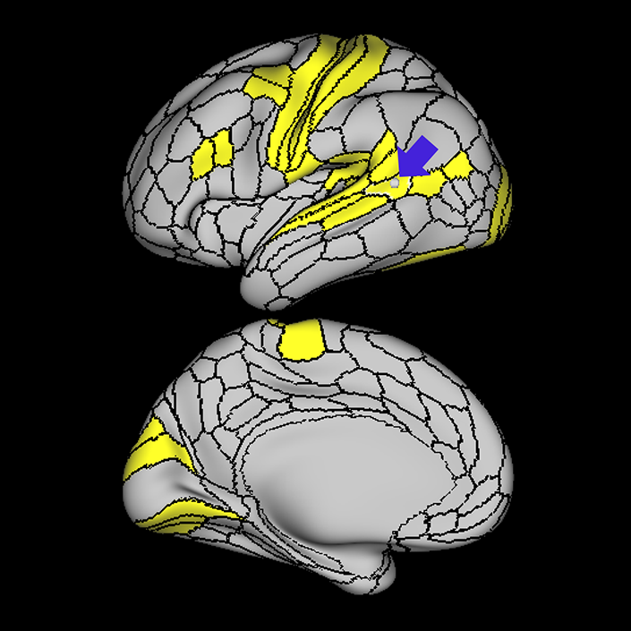
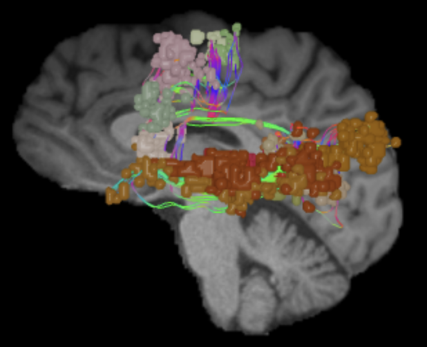
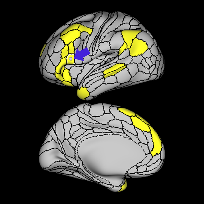
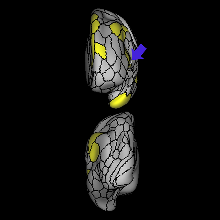
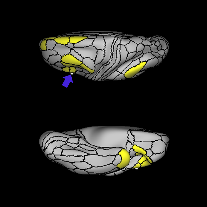
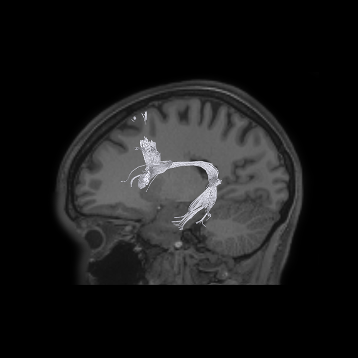
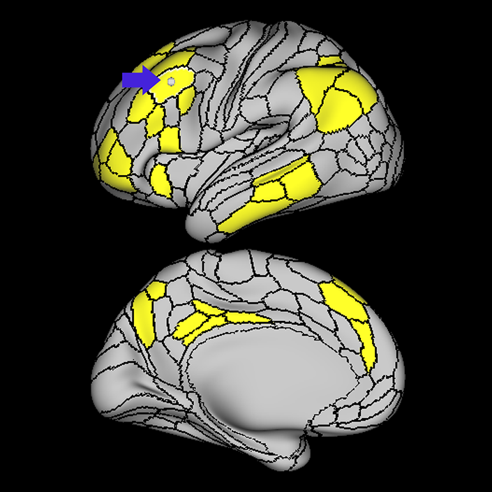
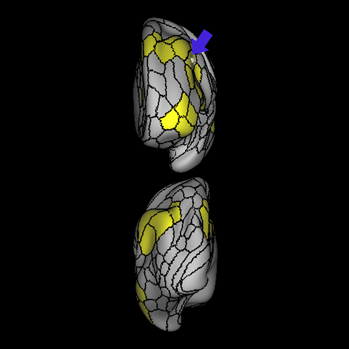
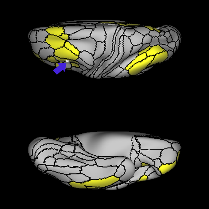
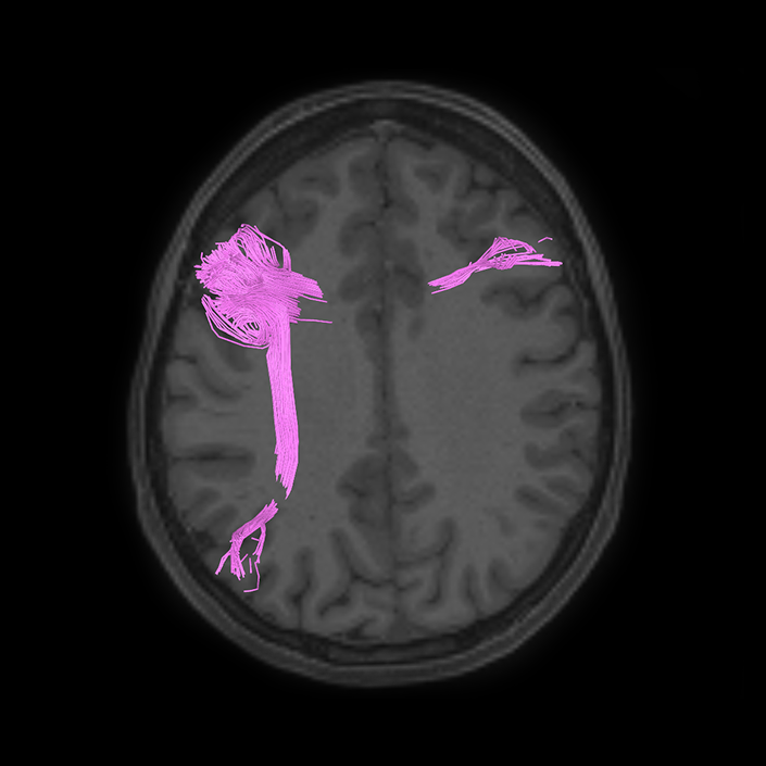
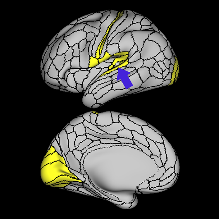
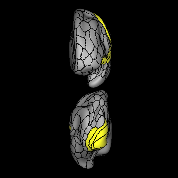
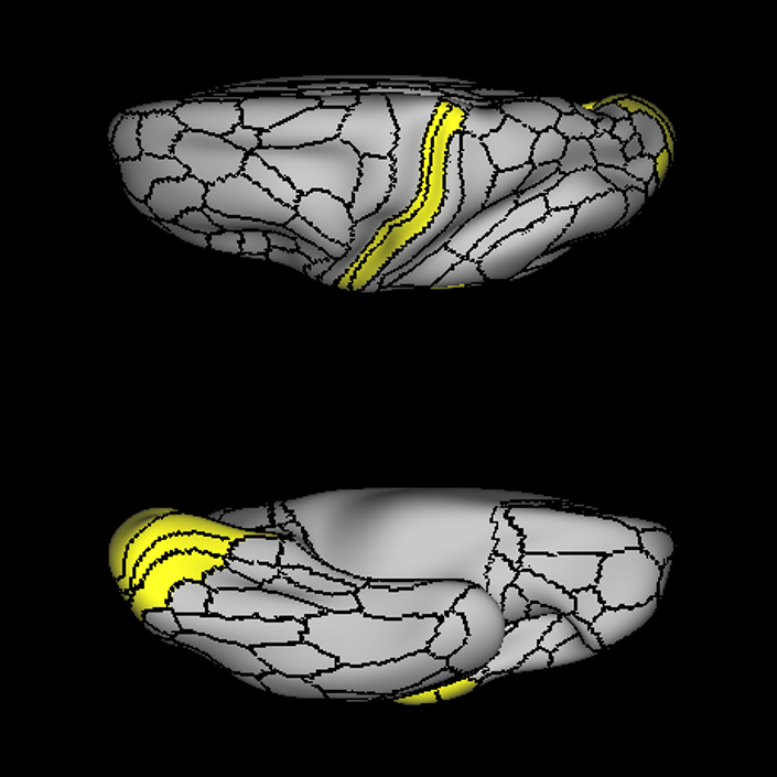
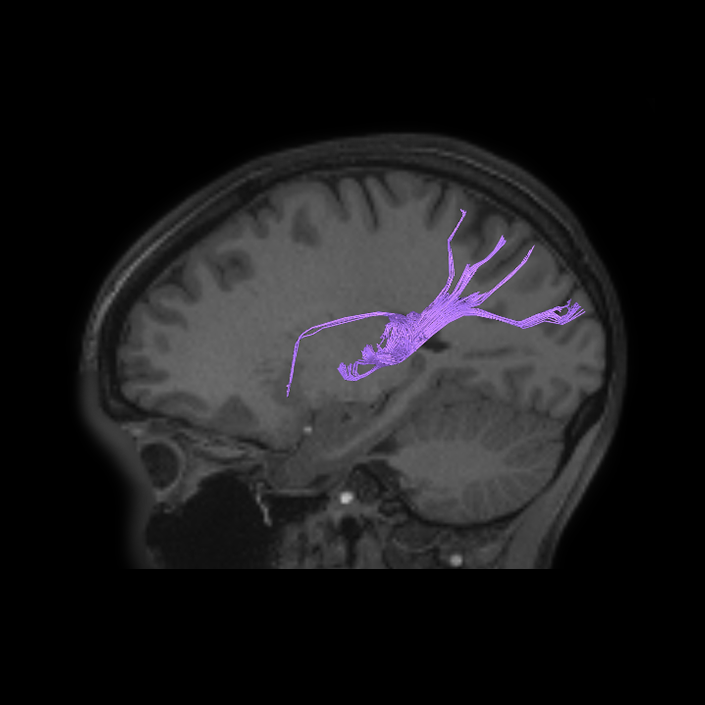
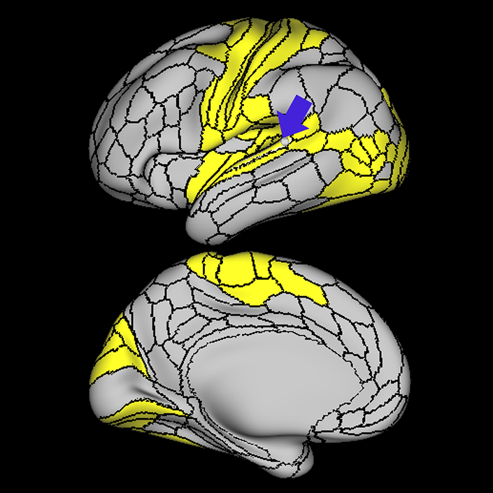
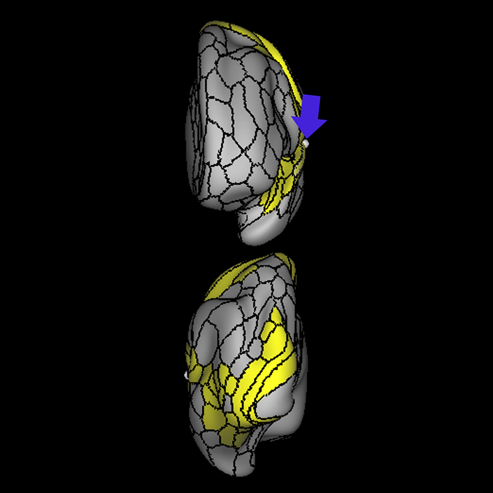
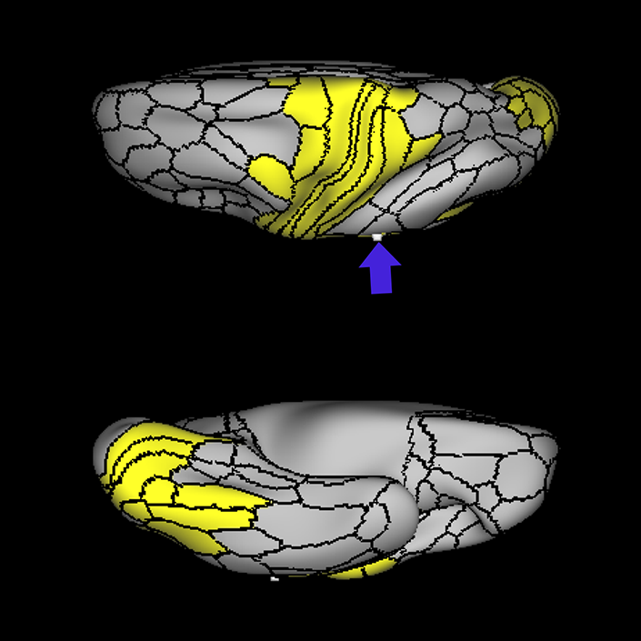
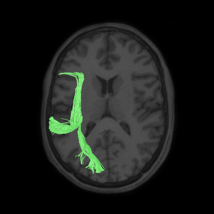
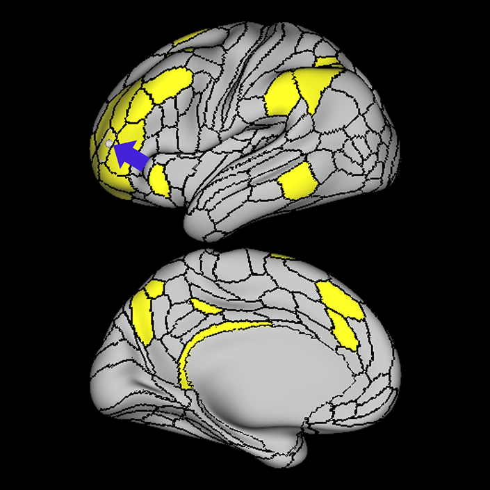
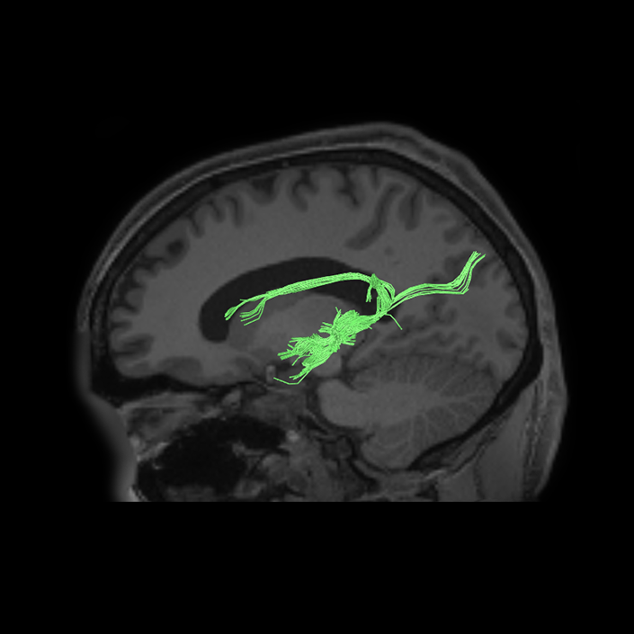
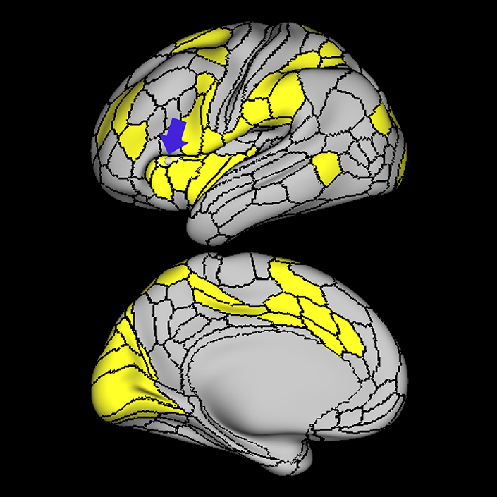
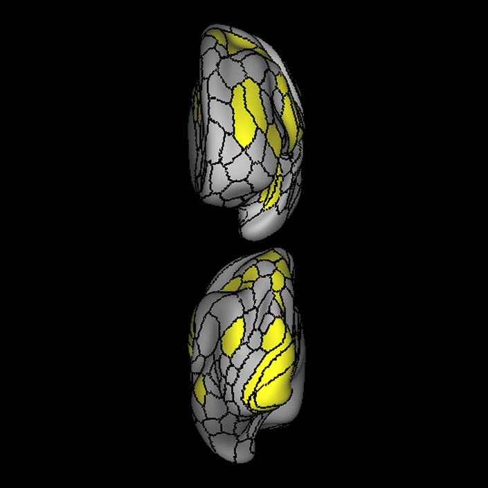
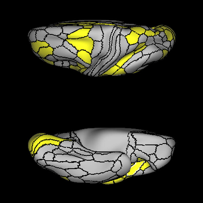
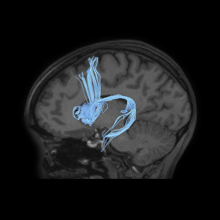
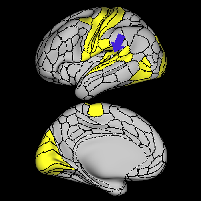
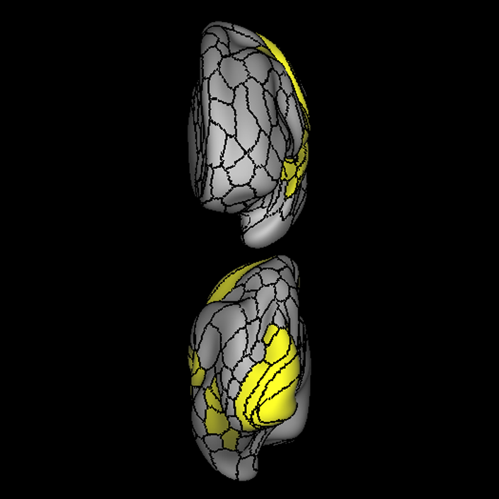
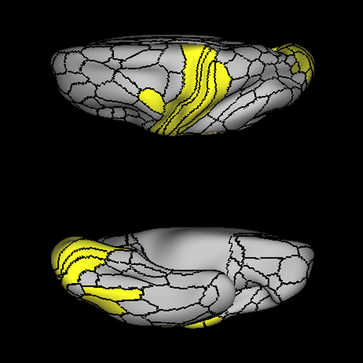
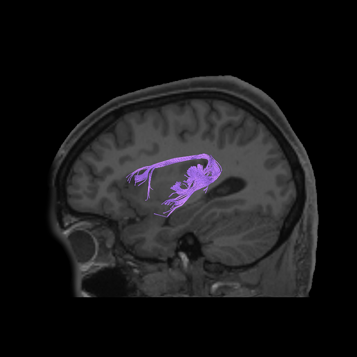
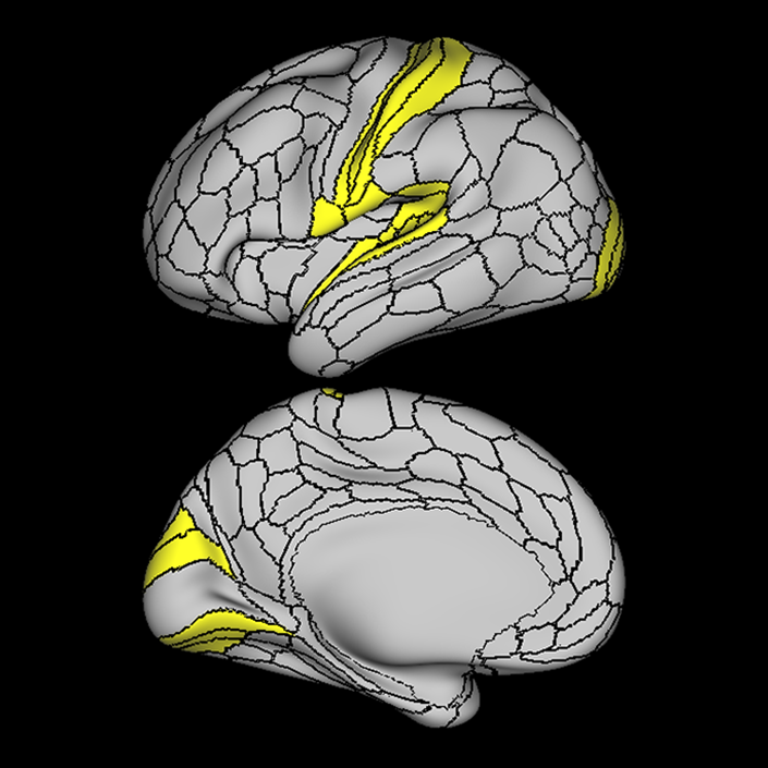
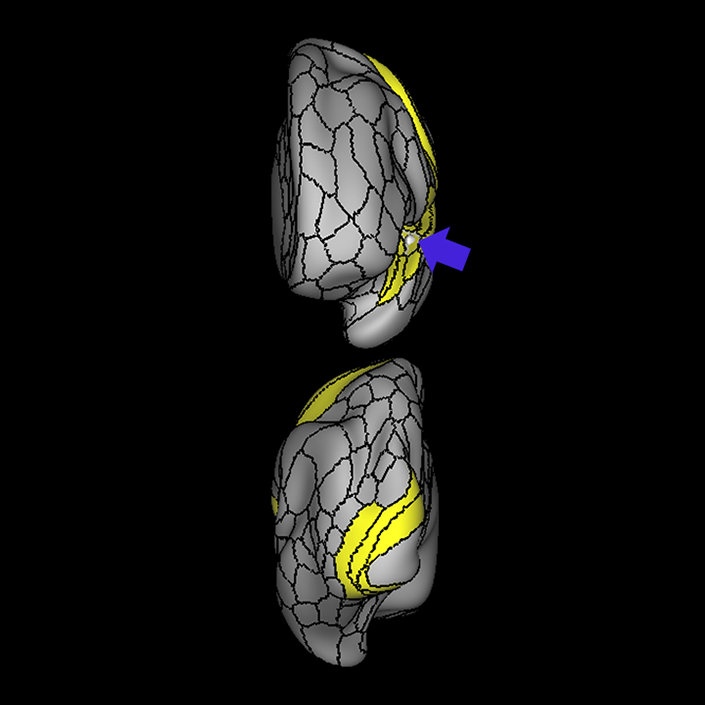
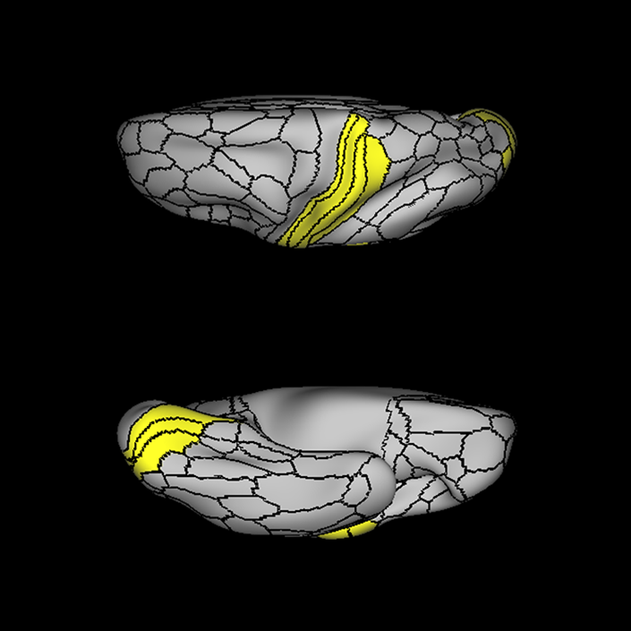
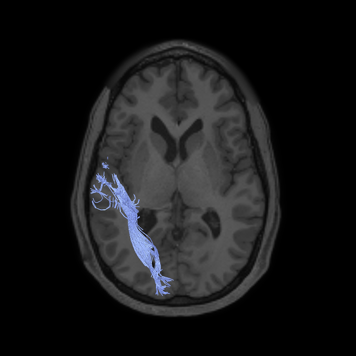
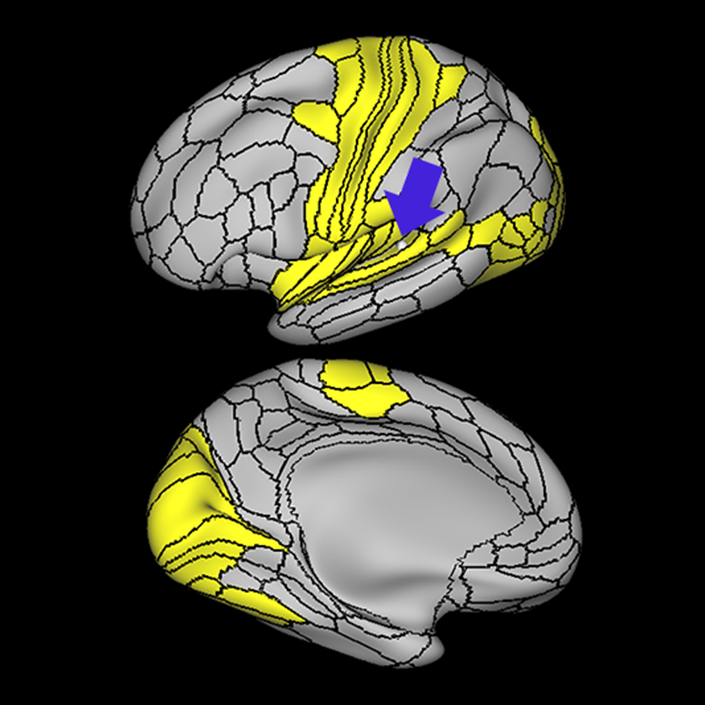
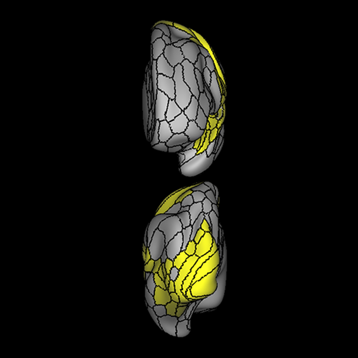
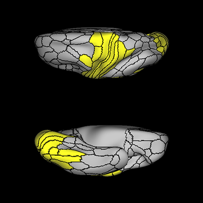
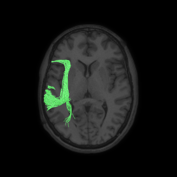
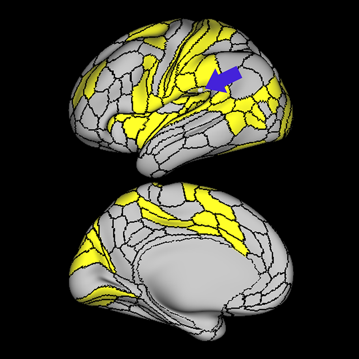
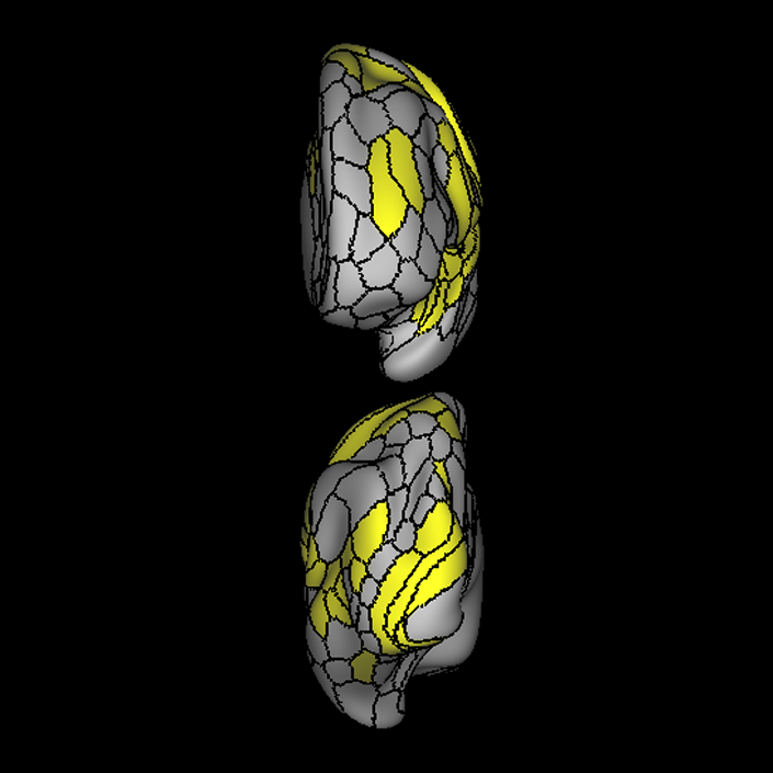
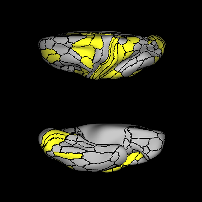
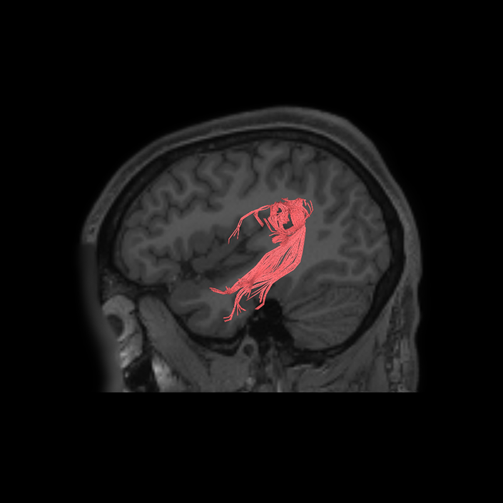
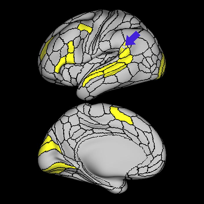
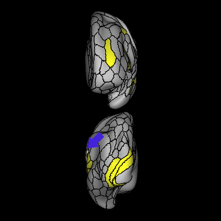
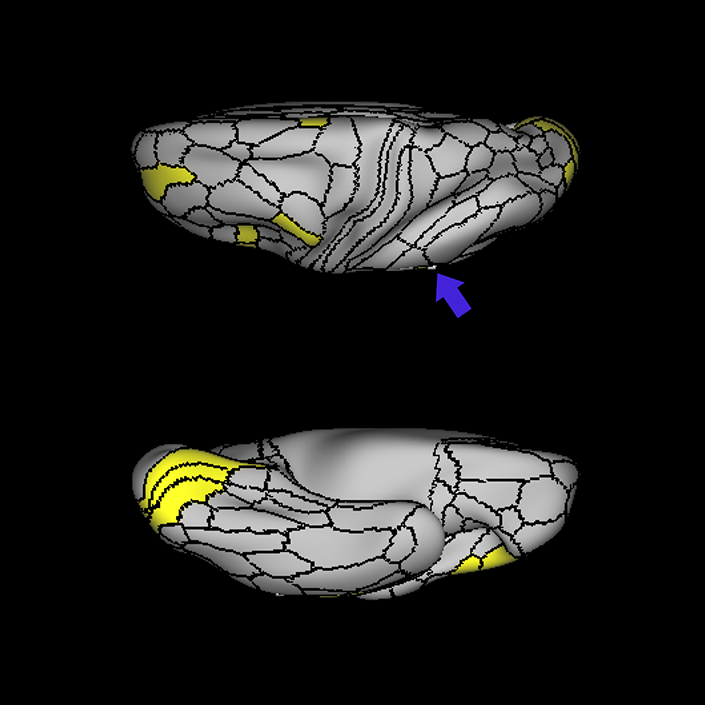
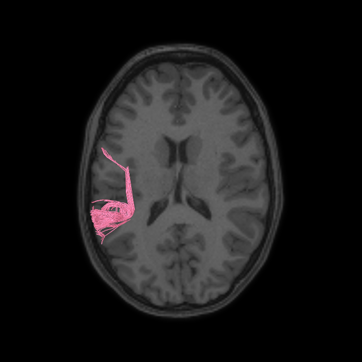
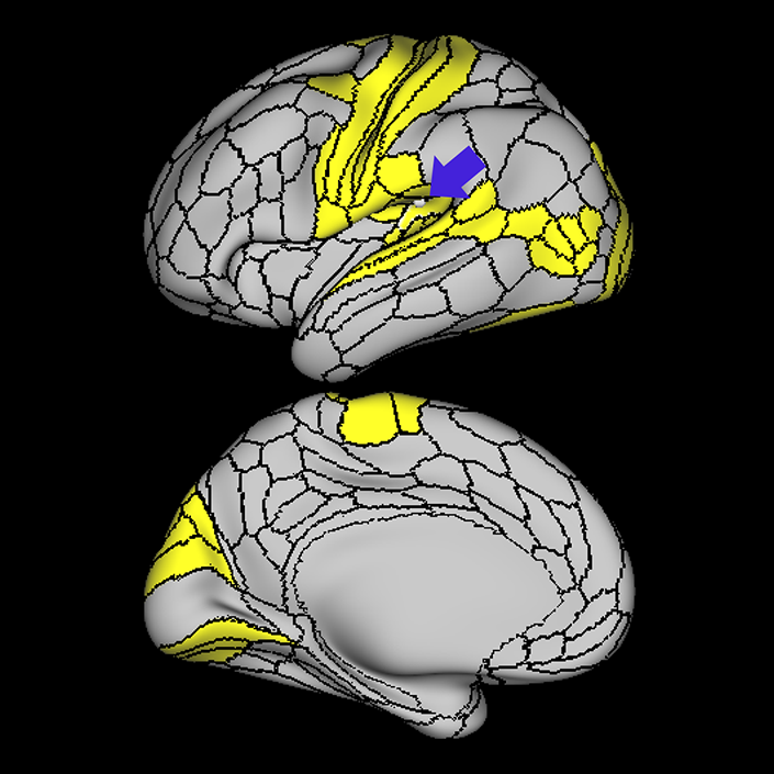
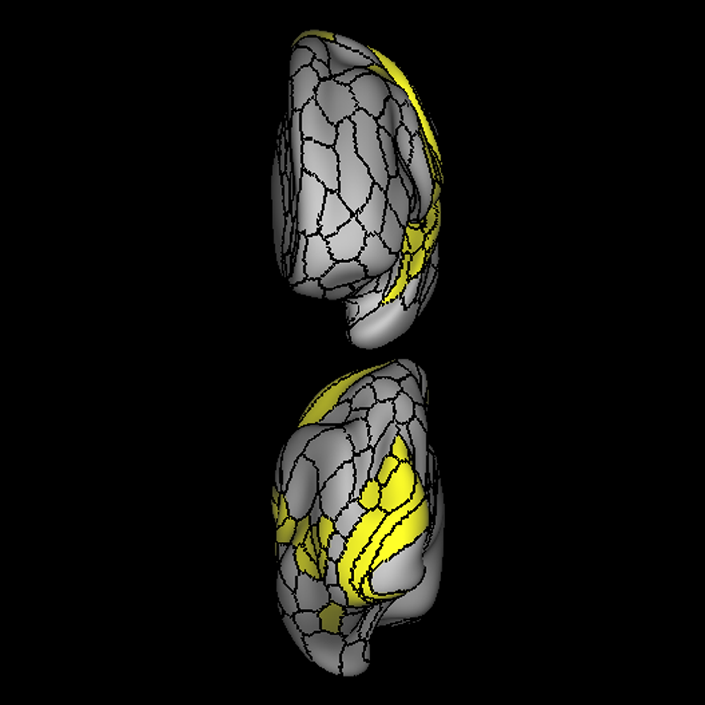
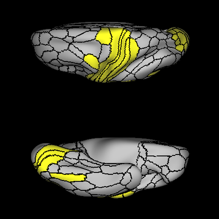
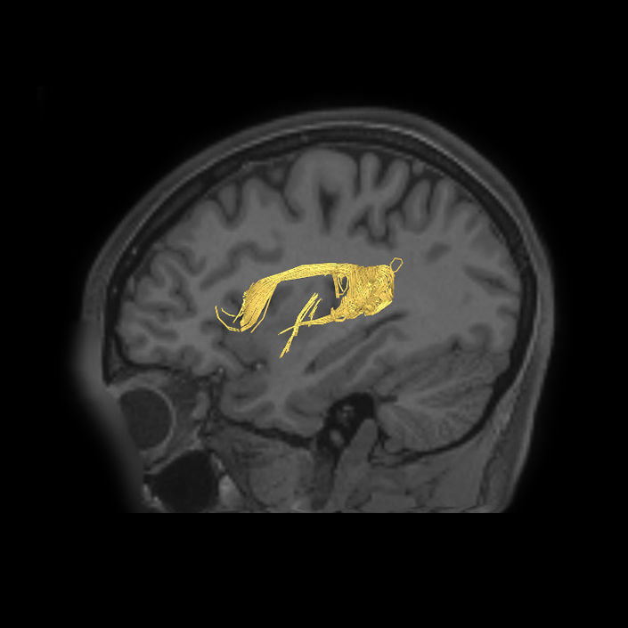
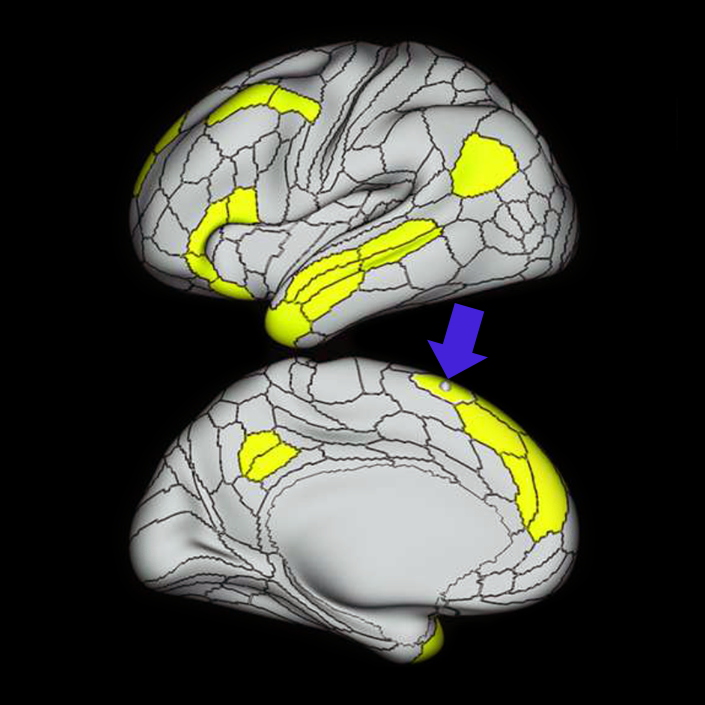
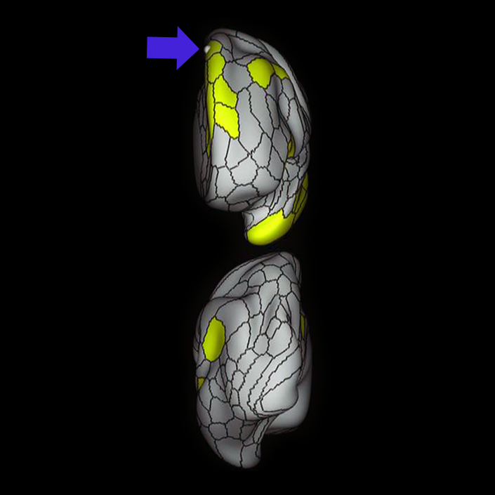
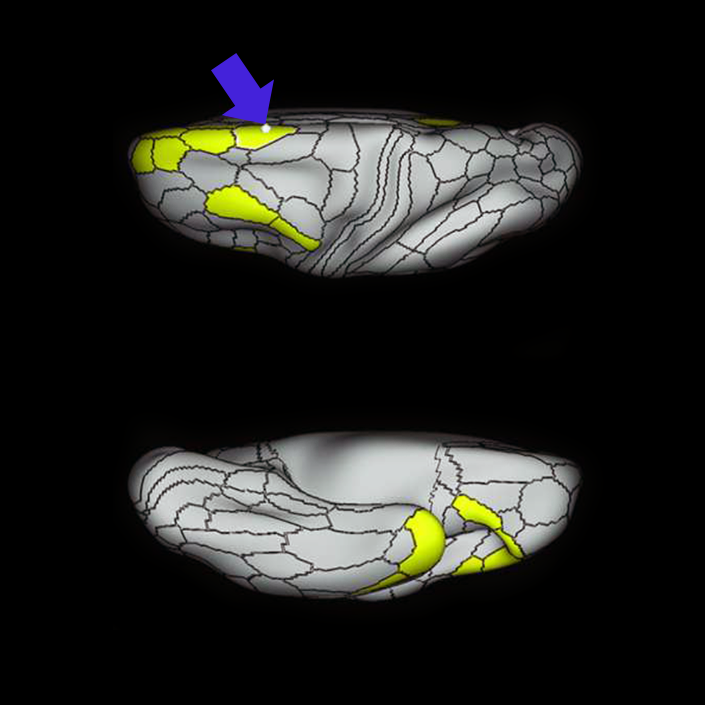
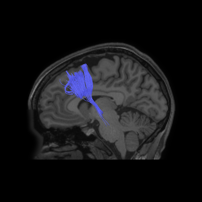
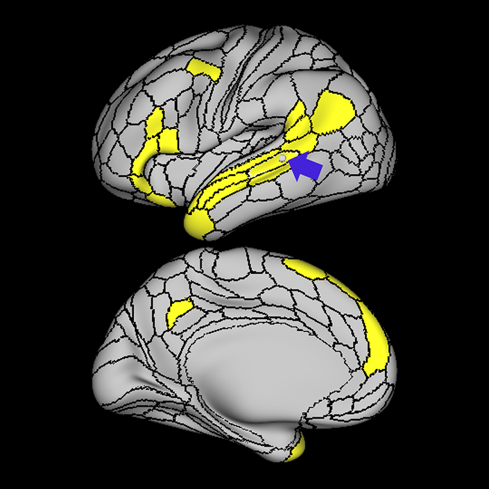
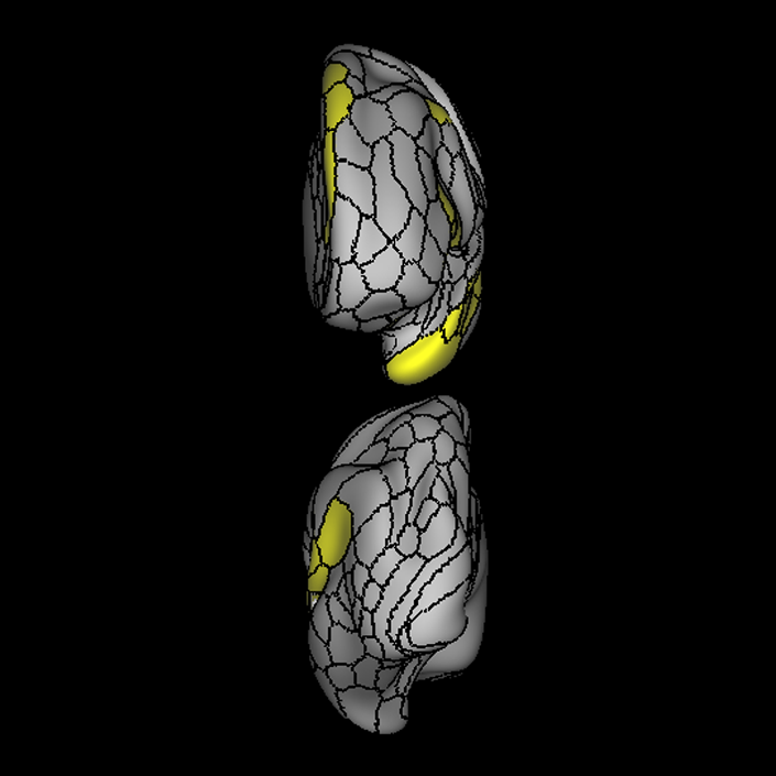
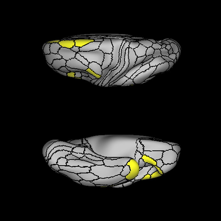
ᐅ SummaryArea 44: part of the inferior frontal gyrus of the lateral frontal lobe. Translates abstract and intentional information in the prefrontal cortex to more detailed representations to help guide the production of verbal and manual actions. In addition to its known association with Broca's area, is sometimes represented as part of Broca's complex ᐅ Where is it?Area 44 is at the posterior most part of the inferior frontal gyrus. It is the anterior bank of pars opercularis of the IFG. ᐅ What are its borders?Area 44 borders area 45 anteriorly and area 6r posteriorly. Area 8C is its medial border and its inferior border is wedged between then upper borders of Areas 6R and 6V. Its superior edge borders IFSp and IFJa. Its opercular surface is FOP4. ᐅ What are its functional connections?Area 44 demonstrates functional connectivity to areas SFL, IFSp, IFJa, 45, 47s, 47L, 9a, 9m, 8AV, 8BL and 8C in the dorsolateral frontal lobe, area 8BM in the medial frontal lobe, area 55b in the premotor areas, areas FOP5, AVI and PSL in the insula- opercular region, areas TGd, STSdp and STSvp in the temporal lobe, areas PFm, and PGi in the inferior parietal lobe, and no areas in the medial parietal lobe. ᐅ What are its white matter connections?Area 44 is structurally connected to the arcuate/SLF and the FAT. Connections with the arcuate/SLF project posteriorly and wrap around the Sylvian fissure to the middle temporal gyrus to end at TE1a and TE1m. There are also projections from the arcuate/SLF before it terminates to parcellations A5 and STSdp. The majority of the inferior connections of the frontal aslant tract end at 44, the tract is connected superiorly to superior frontal gyrus parcellations SFL, 6ma and s6-8. Local short association bundles are connected with 45 and 8C. White matter tracts from 44 in the right hemisphere have less consistent connections with the arcuate/SLF. ᐅ What is known about its function?Area 44 translates abstract and intentional information in the prefrontal cortex to more detailed representations to help guide the production of verbal and manual actions. Area 44, in addition to its known association with Broca's area, is sometimes represented as part of "Broca's complex", including Brodmann Areas 45, 46, 47 and the mesial supplementary motor area of 6, which contribute to a frontal-subcortical circuit. The right pars opercularis has also been implicated in cognitive inhibition in the overall context of working memory. |
|
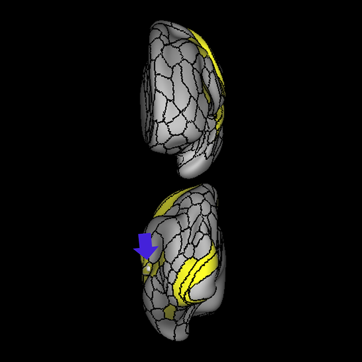
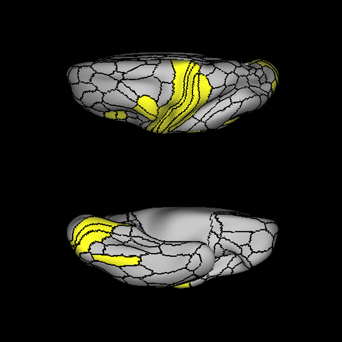
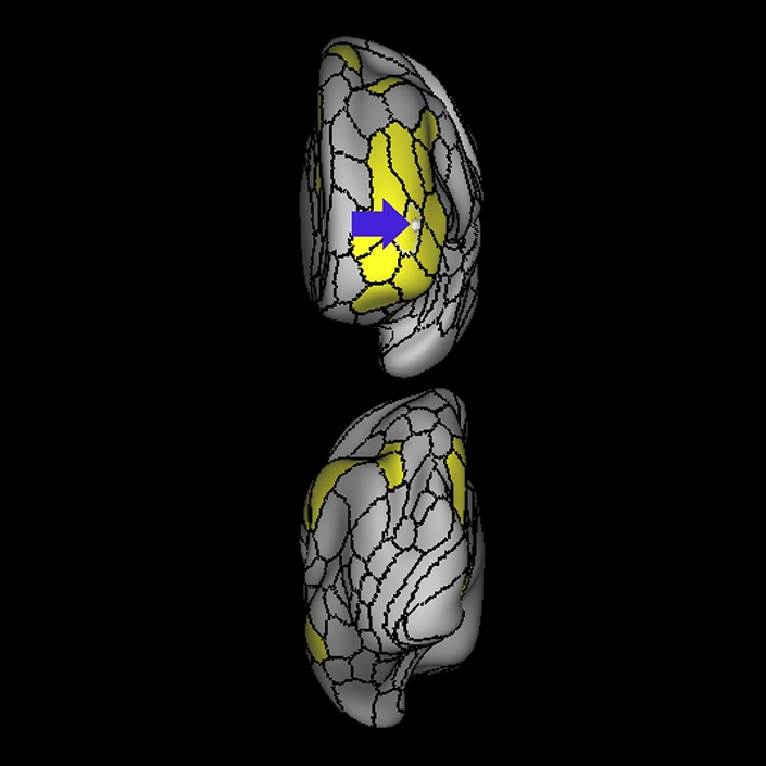
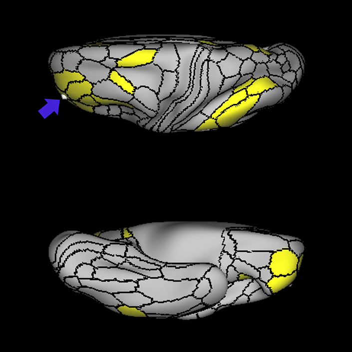
A: lateral-medial
B: anterior-posterior
C: superior-inferior
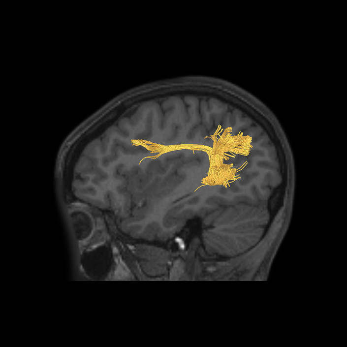
DTI image |
ᐅ SummaryArea 8C: part of the lateral frontal lobe regions. Within the context of spatial working memory, area 8C is involved in the interpretation of complex visual information and attention. Areas 8 and rostral 6, as part of the posterior dorsolateral frontal areas, are also involved in the maintenance of spatial information. ᐅ Where is it?Area 8C is located at the posterior part of the middle frontal gyrus. It is an anterior- to-posterior band which is lateral to area 8AV. ᐅ What are its borders?Area 8C has area 8AV as its main medial border. Its lateral border is with 3 inferior frontal sulcus areas: IFSp, IFJa, and IFJp. Area 55b and PEF (precentral eyefield) are its posterior borders (and discussed in a different section). Area p9-46v and to a lesser extent area 46 are its anterior neighbors. ᐅ What are its functional connections?Area 8C demonstrates functional connectivity to areas s6-8, i6-8, a9-46v, p9-46v, a10p, 8BL, 8AD, and 8AV in the dorsolateral frontal lobe, areas 8BM and d32 in the medial frontal lobe, areas IFSp, IFJp, a47r, p47r, and 44 in the inferior frontal lobe, area AVI in the insula, areas TE1m, TE1p, TE2a, STSva, and STSvp in the temporal lobe, areas IP1, IP2, LIPd, PFm, PGi and PGs in the inferior parietal lobe, and areas 7pm, 31pv, 31a, POS2, 23d, and d23ab in the medial parietal lobe. ᐅ What are its white matter connections?Area 8C is structurally connected to the arcuate/SLF and the contralateral hemisphere. Contralateral connections travel through the corpus callosum to end at 8C. Connections with the arcuate/SLF project posteriorly and wrap around the sylvian fissure to the posterior temporal lobe to end at PH and PHT. ᐅ What is known about its function?Within the context of spatial working memory, area 8C is involved in the interpretation of complex visual information and attention. Areas 8 and rostral 6, as part of the posterior dorsolateral frontal areas, are also involved in the maintenance of spatial information. |
|
A: lateral-medial
B: anterior-posterior
C: superior-inferior
DTI image |
ᐅ SummaryArea A1 (primary auditory cortex): part of parietal apex regions. Receives input from the ventral division of the medial geniculate complex and contains a tonotopic map which then interprets input received from the cochlea. ᐅ Where is it?Area A1 (Primary auditory cortex) is located in superior central portion of Heschl's gyrus. ᐅ What are its borders?Area A1 is largely surrounded by the MBelt and Lbelt. Its deep surface borders the RI area. ᐅ What are its functional connections?Area A1 demonstrates functional connectivity to areas 2, 3a, and 3b in the sensory strip, areas 43, PFcm, and OP4 in the superior opercular region, areas A4, MBelt, PBelt, LBelt, RI, and 52 in the inferior insula opercular region, and areas V1, V2, V3, and V4 in the medial occipital lobe. ᐅ What are its white matter connections?Area A1 is structurally connected to the parietal lobe, occipital lobe, inferior frontal gyrus and local parcellations. Parietal connections are likely portions of the middle longitudinal fasciculus. Fibers from A1 project posterior to parital parcellations LIPv and 7PC, and to occipital parcellations V3B, V3CD and V4. Fibers that project anteriorly to the inferior frontal gyrus connect to 44. Local short association bundles connect with 52, LBelt, MBelt, PBelt and MI. ᐅ What is known about its function?Area A1, also known as the primary auditory cortex, receives input from the ventral division of the medial geniculate complex and contains a tonotopic map which then interprets input received from the cochlea. |
|
A: lateral-medial
B: anterior-posterior
C: superior-inferior
DTI image |
ᐅ SummaryArea A4 (auditory area 4): part of temporal hypotenuse regions. Parcellated from the auditory association cortex. There is evidence that this regions of the brain processes perceptual and conceptual acoustic sounds. ᐅ Where is it?Area A4 (auditory area 4) is on the superior face of the posterior half of the STG. It occupies this portion of the gyrus posterior to its junction with Heschl's gyrus. ᐅ What are its borders?Area A4 borders A5 laterally and PBelt medially. Its posterior borders are with PSL and STV. Its anterior border is with MBelt and TA2. ᐅ What are its functional connections?Area A4 demonstrates functional connectivity to areas 1, 2,3a and 3b in the sensory strip, area 4 in the motor strip, areas SCEF, FEF, 6mp, and 6v in the premotor regions, areas 5m, 5l, and 24dd in the medial frontal lobe, areas 43, PFcm, OP4, OP2-3 and OP1 in the superior insula opercular regions, areas STV, 52, RI, TA2, PI, MI, PBelt, MBelt, LBelt, A1, A5, PoI1, and PoI2 in the lower opercula and Heschl's gyrus regions, areas 7PC and PFop, in the lateral parietal lobe, areas V2, V3, and V4 in the medial occipital lobe, areas V6, V6a, V7, and V3a in the dorsal visual stream area, areas V8, FFC, VVC, and PIT in the ventral visual stream regions, and areas TPOJ1, TPOJ2, LO1, LO3, MT, MST, PH, and FST in the lateral occipital lobe ᐅ What are its white matter connections?Area A4 is structurally connected to the arcuate/SLF and middle longitudinal fasciculus. Arcuate/SLF fibers wrap around the termination of the sylvian fissure from A4 to inferior frontal gyrus parcellations 45 and FOP5. The middle longitudinal fasciculus projects posterior from A4 to run just lateral to the lateral ventricle ending at occipital and parietal lobe parcellations V2, V3, V6, V7, MIP, LIPd, LIPv and IP1. Local short association bundles connect with A5, PFop, RI, STV, LBelt, MBelt, PBelt and PFcm ᐅ What is known about its function?Area A4 is a newly described area of the brain parcellated from the auditory association cortex. There is evidence that this region of the brain processes perceptual and conceptual acoustic sounds. Area A4 was differentiated from area TA2 based on differences in fMRI activity during arithmetic and auditory story tasks. |
|
A: lateral-medial
B: anterior-posterior
C: superior-inferior
DTI image |
ᐅ SummaryArea A5 (auditory area 5): part of temporal hypotenuse regions. There is evidence that this regions of the brain processes perceptual and conceptual acoustic sounds. ᐅ Where is it?Area A5 (Auditory area 5) is a thin anterior-posterior running strip on the superior lateral surface of the posterior portions of the STG. ᐅ What are its borders?Area A5 shares its primary medial border with A4. Its primary posterior border is with TPOJ1. Its lateral borders are with STSdp and STSda (discussed with the temporal lobe section. Its anterior border is with TA2. ᐅ What are its functional connections?Area A5 demonstrates functional connectivity to areas 1, 2, 3a and 3b in the sensory strip, area 4 in the motor strip, area OP4 in the superior insula opercular regions, areas STV, RI, STGa, TA2, PBelt, LBelt, A4, and PSL in the lower opercula and Heschl's gyrus regions, areas STSda, and STSdp in the temporal lobe, area FFC in the ventral visual stream regions, and areas TPOJ1 and MT in the lateral occipital lobe ᐅ What are its white matter connections?Area A5 is structurally connected to the arcuate/SLF and middle longitudinal fasciculus. Arcuate/SLF fibers wrap around the termination of the sylvian fissure from A5 to inferior frontal gyrus parcellation 44. The middle longitudinal fasciculus projects posterior from A5 to run just lateral to the lateral ventricle to end at intraparietal sulcus parcellations V6 and V6A. Local short association bundles connect with A4, STSda and STSdp ᐅ What is known about its function?Area A5 is a newly described area of the brain parcellated from the auditory association cortex. There is evidence that this region of the brain processes perceptual and conceptual acoustic sounds. Area A5 was differentiated from area TA2 based on differences in fMRI activity during arithmetic, auditory story, and social interaction tasks. |
|
A: lateral-medial
B: anterior-posterior
C: superior-inferior
DTI image |
ᐅ SummaryArea FOP4 (frontal operculum 4): part of anterior apex regions. The frontal operculum plays a key role in the initiation of language and lexical retrieval required for language learning. ᐅ Where is it?Area FOP4 (Frontal operculum 4) is located on the inner surface of the pars opercularis of the IFG. ᐅ What are its borders?Area FOP4 borders area 44 on its exterior surface as well as a small border with area 6r posterosuperiorly. Its posterior border is made up of FOP1 and FOP3. Its inferior border is with MI and AVI. Its anterior border is with FOP5. ᐅ What are its functional connections?Area FOP4 demonstrates functional connectivity to areas SCEF, FEF, PEF, 6v, 6a, 6ma, and 6r in the premotor regions, areas IFSa, 46, and 9-46d in the lateral frontal lobe, areas 5mv, 23c, 24dv, a24prime, p24prime, a32prime, and p32prime in the medial frontal lobe, areas 43, PFcm, OP4, FOP3, and FOP5 in the superior insula opercular regions, areas AVI, MI, PI, 52, PoI1, and PoI2 in the lower opercula and Heschl's gyrus regions, area PHT in the temporal lobe, areas 7AL, 7PL, AIP, LIPv, LIPd, PF, PFt, PGp, and PFop in the lateral parietal lobe, area V6 in the dorsal visual stream area, area 7am and DVT in the medial parietal lobe, areas V1, V2, and V3 in the medial occipital lobe. ᐅ What are its white matter connections?Area FOP4 is structurally connected to the frontal aslant tract and the arcuate/SLF. Connections from FOP4 with the frontal aslant tract project superior to the superior frontal gyrus to end at parcellations 6ma and SFL. Arcuate/SLF fibers project posteriorly above the insula, curving around the termination of the sylvain fissure to end at TGd and TE1a. From the arcuate/SLF there are also projections to the superior temporal gyrus that end at A4 and A5. Local short association fibers connect with AVI, FOP3, FOP5 and MI 14. White matter connections from FOP4 in the right hemisphere have less consistent connections with the arcuate/SLF ᐅ What is known about its function?Area FOP4 is a newly described area of the brain that was parcellated from the frontal operculum. The frontal operculum plays a key role in the initiation of language and lexical retrieval required for language learning. Area FOP4 was parcellated from areas AVI and FOP1 based on differences in functional activity in motor-based tasks. |
|
A: lateral-medial
B: anterior-posterior
C: superior-inferior
DTI image |
ᐅ SummaryArea LBelt (lateral belt): part of parietal apex regions. Parcellated from the auditory cortex. ᐅ Where is it?Area LBelt (Lateral belt) is located on the lateral surface of Heschl's Gyrus. ᐅ What are its borders?Area LBelt borders A1 and part of Mbelt medially and Pbelt laterally. Its deep surface abuts RI. ᐅ What are its functional connections?Area LBelt demonstrates functional connectivity to areas 1, 2, 3a, and 3b in the sensory strip, areas 43, PFcm, OP1, and OP4 in the superior opercular region, areas PoI1, PoI2, A1, A4, A5, Mbelt,, MBelt, STV, Ta2, PI, RI, and 52 in the inferior insula opercular region, and areas V1, V2, V3, and V4 in the medial occipital lobe. ᐅ What are its white matter connections?Area LBelt is structurally connected to the arcuate/SLF. Arcuate/SLF fibers wrap around the termination of the sylvain fissure ending at inferior frontal gyrus and insula parcellations 44 and MI. Local short association bundles, include the temporal terminations of the arcuate/SLF to parcellations MBelt, PBelt, A4 and A5. White matter tracts from LBelt in the right hemisphere have more insula projections from the arcuate/SLF. ᐅ What is known about its function?Area LBelt is a newly described area of the brain parcellated from the auditory cortex. Area LBelt was differentiated from area PBelt and RI based on differences in activity on fMRI during arithmetic, auditory story, and social interaction tasks. |
|
A: lateral-medial
B: anterior-posterior
C: superior-inferior
DTI image |
ᐅ SummaryArea MBelt (medial belt): part of parietal apex regions. Parcellated from the auditory cortex. ᐅ Where is it?Area MBelt (Medial belt) runs on the medial surfaces of Heschl's gyrus. ᐅ What are its borders?Area MBelt borders A1 on its lateral surface and IG and area 52 on its medial surface. Its superior edge borders RI and its inferior edge borders TA2. ᐅ What are its functional connections?Area MBelt demonstrates functional connectivity to areas 1, 2, 3a, and 3b in the sensory strip, areas 43, PFcm, and OP4 in the superior opercular region, areas PoI1, A1, A4, Pbelt, LBelt, Ta2, PI, RI, and 52 in the inferior insula opercular region, and areas V1, V2, V3, and V4 in the medial occipital lobe. ᐅ What are its white matter connections?Area MBelt is structurally connected with the middle longitudinal fasciculus. Fibers from the middle longitudinal fasciculus project posterior just lateral to the lateral ventricle to occipital and parietal lobes to end at parcellations V3, V3A, V6, V6A, IPS1 and DVT. The majority of short association bundles connect anterior to A4, TA2, STGa, and locally to PBelt and LBelt. ᐅ What is known about its function?Area MBelt is a newly described area of the brain parcellated from the auditory cortex. Area MBelt was differentiated from area 52 based on differences in activity on fMRI during arithmetic and auditory story tasks. In addition, MBelt was differentiated from PBelt based on differences in activity on fMRI during arithmetic, auditory story, and object recognition tasks. |
|
A: lateral-medial
B: anterior-posterior
C: superior-inferior
DTI image |
ᐅ SummaryArea PBelt (parabelt complex): part of parietal apex regions and superior insula opercular regions. Parcellated from the auditory cortex. ᐅ Where is it?Area PBelt (Parabelt complex) is located on the superior surface of the inferior portion of the supramargina gyrus. It lies in the small region between the lateral edge of Heschl's gyrus and the opercular cleft of the inferior SMG. ᐅ What are its borders?Area PBelt borders Lbelt and Mbelt medially and A4 laterally. Its deep surface borders with RI ᐅ What are its functional connections?Area PBelt demonstrates functional connectivity to areas 1, 2, 3a, and 3b in the sensory strip, area 4 in the motor strip, area 24dd in the paracingulate areas, areas FEF, 6d, 6v, and 6mp in the premotor areas, areas 43, PFcm, OP1, OP2-3, and OP4 in the superior opercular region, areas A1, A4, A5 Mbelt, LBelt, PoI1, PoI2, STV, Ta2, PI, RI, and 52 in the inferior insula opercular region, areas PFop and 7PC in the parietal lobe, areas V1, V2, V3, and V4 in the medial occipital lobe, areas V6, V67a, V7, V3a, and V3b in the dorsal visual stream, areas V8, FFC, Pit and VVC in the ventral visual stream, and areas LO2, LO3, V3cd, FST, MT, MST, V4t, TPOJ1, and TPOJ2 in the lateral occipital lobe. ᐅ What are its white matter connections?Area PBelt is structurally connected to the middle longitudinal fasciculus and arcuate/SLF. Arcuate/SLF fibers wrap around the termination of the sylvian fissure to end at the inferior frontal gyrus at parcellation 45 and FOP5. Fibers from the middle longitudinal fasciculus project posterior just lateral to the lateral ventricle to occipital and parietal lobes to end at parcellations MIP, LIPv and IP1. Local short association bundles connect with A1, LBelt, MBelt, PFcm, PSL, A4, A5, TPOJ1. ᐅ What is known about its function?Area PBelt is a newly described area of the brain parcellated from the auditory cortex. Area PBelt was differentiated from areas A4 and LBelt based on differences in activity on fMRI during arithmetic, auditory story, and motor cue tasks. |
|
A: lateral-medial
B: anterior-posterior
C: superior-inferior
DTI image |
ᐅ SummaryArea PFcm (parietal F, regions cm): part of parietal apex regions, and subdivision of the inferior parietal cortex. The inferior parietal cortex is believed to be important in processing language with regard to language vocabulary, semantics, articulation, and working memory. ᐅ Where is it?Area PFcm (Parietal F, region cm) is located in the superior portion of the supramargina gyrus. It is primarily located on the opercular cleft of this part of the gyrus What are its borders? Area PFcm borders borders parietal regions PF, and PFop superiorly. Its anteroinferior border is OP1. RI is its inferior border. Its posterior border is PSL. ᐅ What are its borders?
ᐅ What are its functional connections?Area PFcm demonstrates functional connectivity to Area OP4 demonstrates functional connectivity to areas 1, 2, 3a, 3b in the sensory strip, areas SCEF, FEF, PEF, 6mp, 6r, 6a and 6v in the premotor regions, areas 24dv, a24prime, p24prime, p32prime, 24dd, 24dv, 5mv, and 23c in the middle cingulate regions, areas 9-46d and 46 in the lateral frontal lobe, areas 43, OP4 OP2-3, OP1, PFcm, FOP1, FOP2, FOP3, FOP4 and FOP5 in the superior insula opercular regions, areas LBelt, PBelt, MBelt, A1, TA2, PI, A4, TA2, MI, STV, 52, RI, PoI1 and PoI2 in the lower opercula area and Heschl's gyrus regions, PHT in the temporal lobe, areas PF, PFop, PFt, PGp, AIP, 7PC, and 7AL in the lateral parietal lobe, areas 7am and DVT in the medial parietal lobe, areas V2 and V3 in the medial occipital lobe, area V3a of the dorsal visual stream, areas FFC of the ventral visual stream, and areas LO3, TPOJ1, TPOJ2, TPOJ3, MST, and FST of the lateral occipital lobe. ᐅ What are its white matter connections?Area PFcm is structurally connected to portions of the arcuate/SLF. Arcuate/SLF connection wrap around the termination of the sylvian fissure with inferior projections through the temporal lobe to end at inferior and middle temporal gyrus parcellations TE1a, STSva and TE2a. Local short association bundles are connected with OP1, OP4, PFop, PBelt, PF and RI. There are less consistent inferior connections from PFcm in the right hemisphere through the temporal lobe. ᐅ What is known about its function?Area PFcm is a subdivision of the inferior parietal cortex. The inferior parietal cortex is believed to be important in processing language with regards to language vocabulary, semantics, articulation and working memory. Area PFcm was differentiated from area PSL based on lower levels of activity on fMRI during arithemetic and auditory story tasks. Area PFcm was also differentiated from area RI based on higher levels of activity on fMRI during motor cue tasks. |
|
A: lateral-medial
B: anterior-posterior
C: superior-inferior
DTI image |
ᐅ SummaryArea PSL (perisylvian language area): part of parietal apex regions. Thought to play a role in higher cognitive functions such as essential information processing, motional control, and control of cognitive functions. Also believed to be associated with special human cognitive functions such as generation of language, visuospatial attention, and assimilation of audiovisual information. ᐅ Where is it?Area PSL (periSylvian Language area) is located in the supramarginal gyrus. It is located at the apex of the posterior Sylvian Fissure, in the lower portion of this posterior part of the SMG. ᐅ What are its borders?Area PSL borders STV inferiorly, PFm posteriorly, PF superiorly, and PFcm anteriorly. It borders RI on its internal surface. ᐅ What are its functional connections?Area PSL demonstrates functional connectivity to area SCEF in the paracingulate areas, area 55b in the premotor areas, areas IFJa, 9-46d, 44 and 45 in the lateral frontal lobe, areas STV and A5 in the inferior insula opercular region, areas STSda, STSdp, and STSvp in the temporal lobe, areas V2, V3, and V4 in the medial occipital lobe, areas V6, V67a, V7, V3a, and V3b in the dorsal visual stream, areas V8, FFC, Pit and VVC in the ventral visual stream, and area TPOJ1 in the lateral occipital lobe. ᐅ What are its white matter connections?Area PSL is structurally connected to the arcuate/SLF. Arcuate/SLF fibers project anteriorly from PSL above the insula to end at 6r, and inferiorly from PSL through the temporal lobe to end at TE1a, STSdp, STSva and STSvp. Local short association bundles connect with PBelt, STV, RI, A5 and PF. ᐅ What is known about its function?Area PSL is a newly described area of the brain parcellated from the temporo-parieto-occipital junction (TPOJ). This area of the brain is thought to play a role in higher cognitive functions such as essential information processing, motional control, and control of cognitive functions. The TPOJ is also believed to be associated with special human cognitive functions such as generation of language, visuospatial attention and assimilation of audiovisual information. Area PSL was differentiated from areas PFcm and RI based on differences in fMRI activity during arithmetic and auditory story tasks. |
|
A: lateral-medial
B: anterior-posterior
C: superior-inferior
DTI image |
ᐅ SummaryArea RI (retroinsular cortex): part of parietal apex regions, and an area of the early auditory cortex. Area RI is connected to various somatosensory areas and receives somatosensory input. As a result, it is believed that this regions plays a role in receiving auditory-somatosensory communications from the auditory cortex in conjunction with the granular insula. ᐅ Where is it?Area RI (Retroinsular cortex) is located in the retroinsular cortex, which itself is located anterior superior to the long gyri of the insula. It is located at the deep termination of Heschl's gyrus as its superomedially. ᐅ What are its borders?Area RI is slanted from anterior-inferior to posterior superior. On its anterior inferior border, it meets IG, OP2-3 and OP1. On its exterior border, it meets PFcm and PSL. On its deep inferoposterior border, it joins with A1, Mbelt, Lbelt, and Pbelt, the Heschl's gyrus regions. ᐅ What are its functional connections?Area RI demonstrates functional connectivity to areas 1, 2, 3a, 3b in the sensory strip, area 4 in the motor strip, areas FEF, 6mp, and 6v in the premotor regions, areas 43, OP4 OP2-3, OP1, PFcm in the superior insula opercular regions, areas LBelt, PBelt, MBelt, A1, TA2, PI, A4, A5, TA2, STV, and 52 in the lower opercula area and Heschl's gyrus regions, area PFop in the lateral parietal lobe, areas V2, V3, and V4 in the medial occipital lobe, area V6, V3b, V3a, and V7 of the dorsal visual stream, areas FFC of the ventral visual stream, and areas LO3, TPOJ1, TPOJ2, TPOJ3, MT, MST V4t, and FST of the lateral occipital lobe. ᐅ What are its white matter connections?Area RI is structurally connected to portions of the arcuate/SLF, anterior insula and local parcellations. Arcuate/SLF fibers project anterior form RI to inferior frontal gyrus parcellations 44 and 6r. Insular fibers course anterior to insular parcellations PoI2 and PoI1. Local short association bundles are connected with A1, OP1, OP4, PBelt and A4 . ᐅ What is known about its function?Area RI is an area of the early auditory cortex. Area RI is connected to various somatosensory areas and receives somatosensory input. As a result, it is believed that this region plays a role in receiving auditory-somatosensory communications from the auditory cortex in conjunction with the granular insula. |
|
A: lateral-medial
B: anterior-posterior
C: superior-inferior
DTI image |
ᐅ SummaryArea SCEF (supplementary and cingulate eye field): part of medial superior frontal gyrus regions. Higher order oculomotor center implicated in appraising all possible oculomotor behaviors to direct primary oculomotor centers in goal- directed behavior. ᐅ Where is it?Area SCEF (supplementary and cingulate eye field) is located in the posterior medial SFG. ᐅ What are its borders?Area SCEF borders area 8BM anteriorly, areas 6ma and SFL superiorly, areas 6mp and 24dd posteriorly, and areas 24dv and p32pr inferiorly. ᐅ What are its functional connections?Area SCEF demonstrates functional connectivity to areas 1, 2, 3a, 3b in the sensory strip, area 4 in the motor strip, areas PEF, FEF, 55b, 6ma, 6mp, 6a, 6r, and 6v in the premotor regions, areas a24prime, p32prime, a32prime, 5mv, and 23c in the middle cingulate regions, areas IFJa, 46, and 9-46d in the lateral frontal lobe areas OP4, OP1, PFcm, 43, FOP1, FOP2, FOP3 FOP4, and FOP5 in the superior insula opercular regions, areas PSL, 52, A4, MI, PoI1 and PoI2 in the lower opercula and Heschl's gyrus regions, area PHT in the temporal lobe, areas AIP, MIP, VIP, LIPd, LIPv, PFop, PF, PFt, IP0, IPS1, 7AL, 7PL, and 7PC, in the lateral parietal lobe, areas 7am and DVT in the medial parietal lobe, area V1, V2, V3, V4 in the medial occipital lobe, areas V3a, V3b, V6, V6a, and V7 in the dorsal visual stream, area FFC of the ventral visual stream, and areas PH, TPOJ2, LO3, MST, and FST of the lateral occipital lobe. ᐅ What are its white matter connections?Area SCEF is structurally connected to the contralateral hemisphere and thalamus. Contralateral connections course through the body of the corpus callosum to end at SCEF, 8BL, SFL and 8BM. Thalamic projections travel through the ventral thalamus to the brainstem (Figure 32). Local short association bundles connect with SF, 8BM, SFL and 8BL. ᐅ What is known about its function?Area SCEF is a higher order oculomotor center implicated in appraising all possible oculomotor behaviors to direct primary oculomotor centers in goal-directed behavior. |
|
A: lateral-medial
B: anterior-posterior
C: superior-inferior
DTI image |
ᐅ SummaryArea STSdp (superior temporal sulcus dorsal posterior): part of the temporal lobe regions. Involved in motion processing, audiovisual integration, and facial processing. The posterior half of STSdp (as with STSvp) is strongly activated in the story-math secondary contrast, indicating a role in language comprehension. STSdp responds more strongly than STSvp to primary language tasks and to social cognition and motor tasks. ᐅ Where is it?Area STSdp (superior temporal sulcus dorsal posterior) is found on the posterior half of the lateral face of the STG and the posterior half of the superior bank of the superior temporal sulcus ᐅ What are its borders?Area STSdp borders area STSda anteriorly, STSvp inferiorly, TPOJ1 posteriorly, and A5 superiorly. ᐅ What are its functional connections?Area STSdp demonstrates functional connectivity to areas 9m, 8BL, 44 45, 47L, 47s, IFSp, SFL and 55b in the frontal lobe, areas STV, PSL, A5, and STGa in the insula opercular area, areas STSva, STSvp, STSda, and TGd, in the temporal lobe, TPOJ1 in the lateral occipital lobe, and PGi and 31pd in the parietal lobe ᐅ What are its white matter connections?Area STSdp is structurally connected to the "u" fibers of the occipito-temporal system and the arcuate/SLF. Arcuate/SLF tracts wrap around the Sylvian fissure projecting toward the frontal lobe and turn medially to terminate at 44, FOP4, IFJa, IFJp and IFSp. Local short association fibers include "u" fibers of the occipito-temrporal system that connect to STSda, STSva, STSvp, PSL and P ᐅ What is known about its function?The posterior portion of the STS is primarily involved in motion processing, audiovisual integration, and facial processing. The posterior half of STSdp (like the posterior half of STSvp) is strongly activated in the story-math secondary contrast, indicating a role in language comprehension. STSdp responds more strongly than STSvp to primary language tasks and to social cognition and motor tasks. |
|
A: lateral-medial
B: anterior-posterior
C: superior-inferior
DTI image |

ᐅ SummaryArea TPOJ1 (temporo-parieto-occipital junction 1): part of the lateral parietal lobe regions. Participates in several processes including detecting incongruities between expected and presented stimuli, reward processing, and saliency. Also shows prominence in theory of mind, and the processing and detection of incongruous concepts and their subsequent resolution. Has specifically been shown to be activated during self-related processing, and receives input from different sensory afferent neurons. ᐅ Where is it?Area TPOJ1 (temporal-parietal-occipital junction 1) is found in the posterior superior temporal sulcus, just as it angles upward to indent the angular gyrus. It makes up both banks and the depth of this portion of the sulcus. ᐅ What are its borders?Area TPOJ1 borders STV superiorly, and PHT inferiorly. Its anterior border is small but borders STSdp, STSvp, A4, and A5. It borders PGi posterosuperiorly and TPOJ2 posteriorly. ᐅ What are its functional connections?Area TPOJ1 demonstrates functional connectivity to areas 1,2, 3a, and 3b in the sensory strip, area 4 in the motor strip, areas 55b and FEF in the premotor areas, areas IFJa and IFSp in the lateral frontal lobe, areas OP4, PFcm, RI, STV, PSL, A4, A5, PBelt, and LBelt in the insula opercular region, area STSdp in the temporal lobe, areas V2, V3, and V4 in the medial occipital lobe, area FFC in the ventral visual stream, and areas TPOJ2 and TPOJ3 in the lateral occipital lobe . ᐅ What are its white matter connections?Area TPOJ1 is structurally connected to the arcuate/SLF and inferior parietal lobe. Arcuate/SLF connections wrap around the sylvian fissure toward the fronal lobe to end at premotor parcellations IFJa, IFJp and 6r. From the arcuate/SLF there are also connections to the inferior parietal lobule that end at PFm, PGi and PGs. Local short association fibers connect with TPOJ2, STSvp and STSdp. ᐅ What is known about its function?The temporal-parietal-occipital junction participates in several processes including detecting incongruities between expected and presented stimuli, reward processing, and saliency. This region also shows prominence in theory of mind, and the processing and detection of incongruous concepts and their subsequent resolution. The TPOJ region has specifically been shown to be activated during self-related processing, and receives input from different sensory afferent neurons. The temporal-parietal-occipital junction is not finely parcellated in the current literature, thus task-based fMRI differences are the only way to delineate their individual functions. |
|
A: lateral-medial
B: anterior-posterior
C: superior-inferior
DTI image |