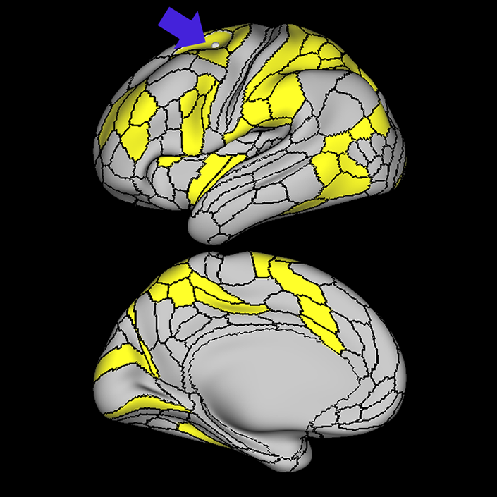
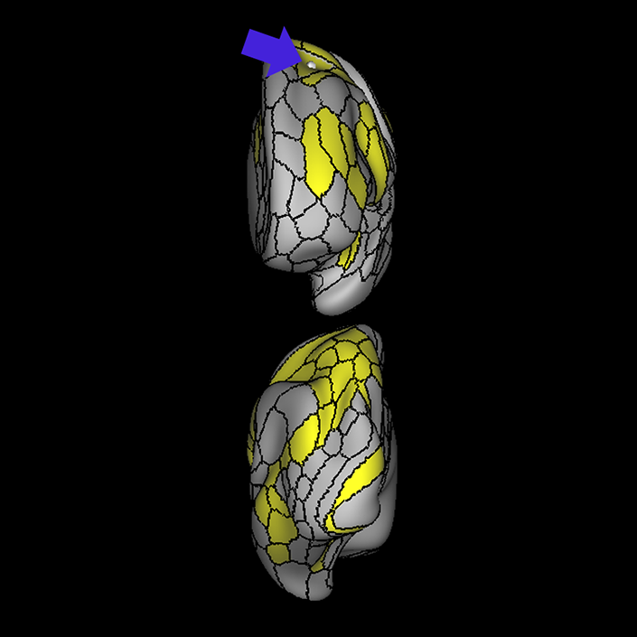
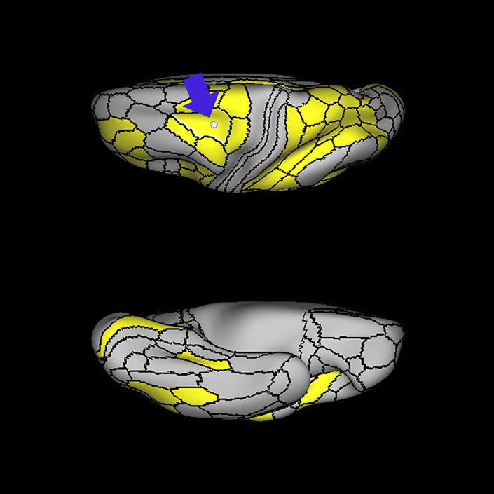
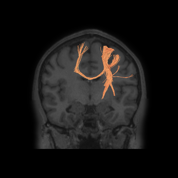
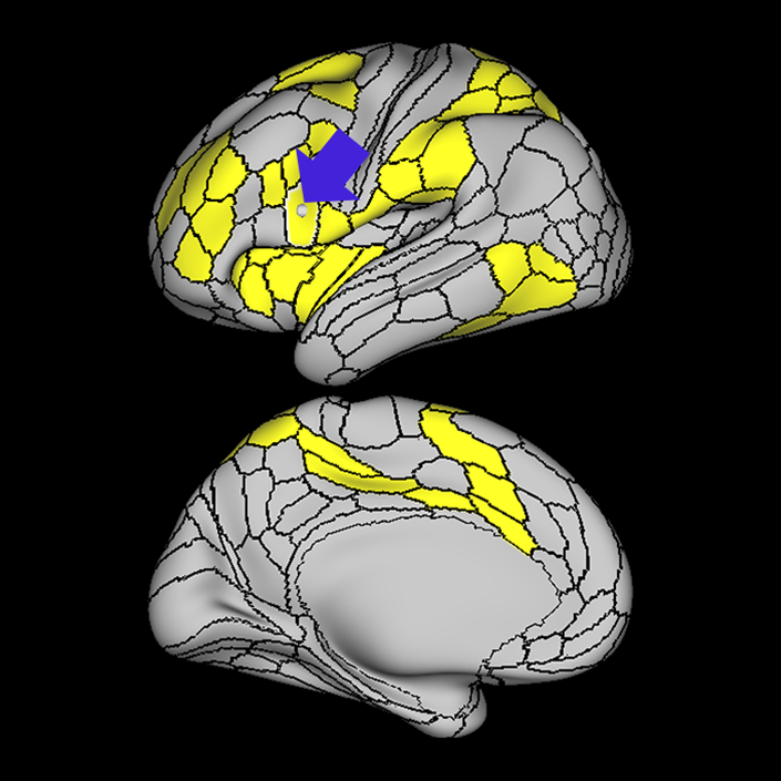
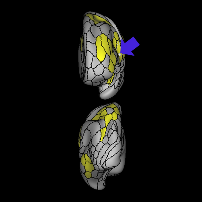
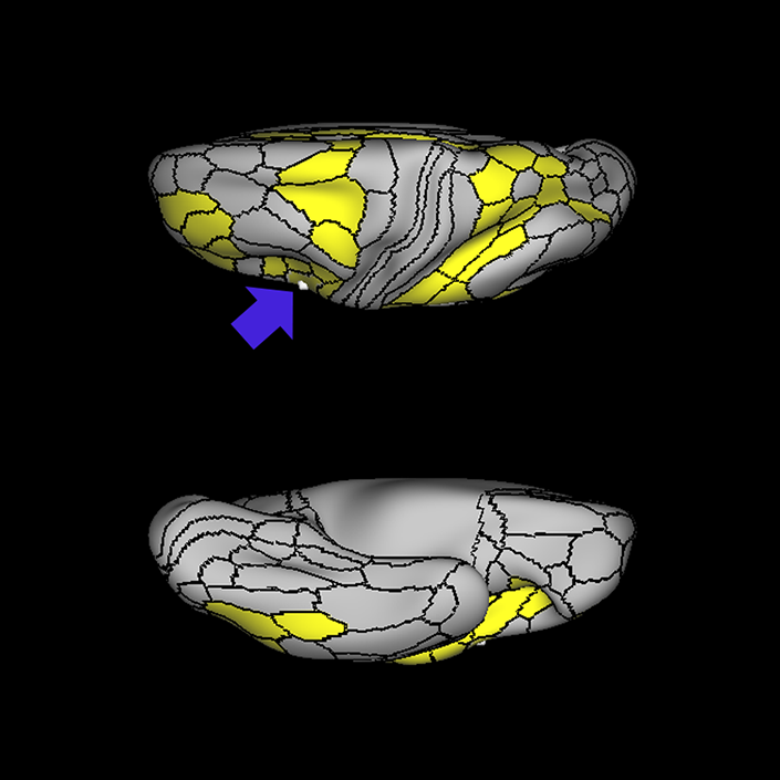
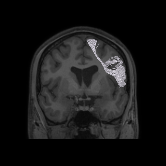
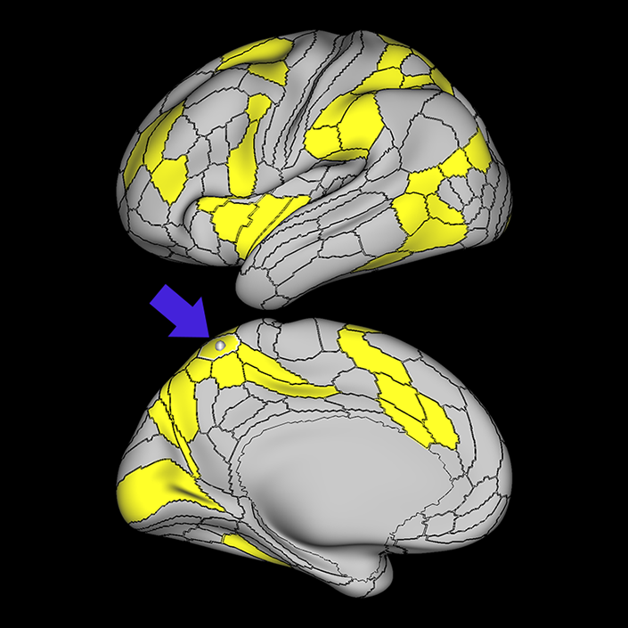
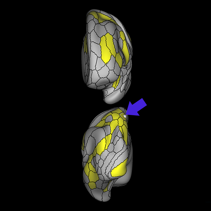
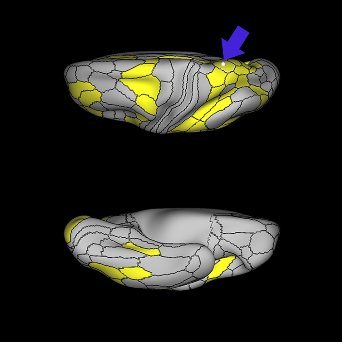
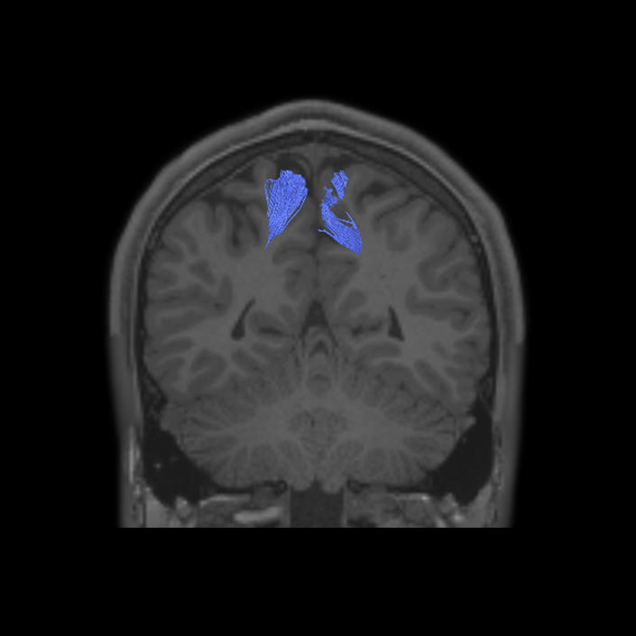
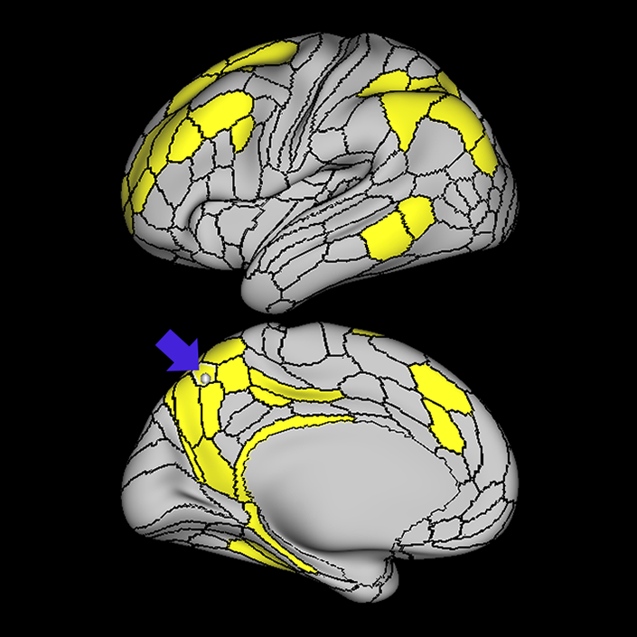
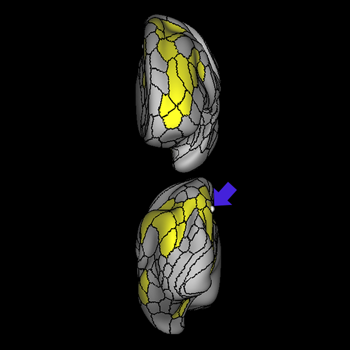
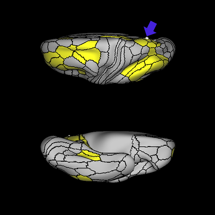
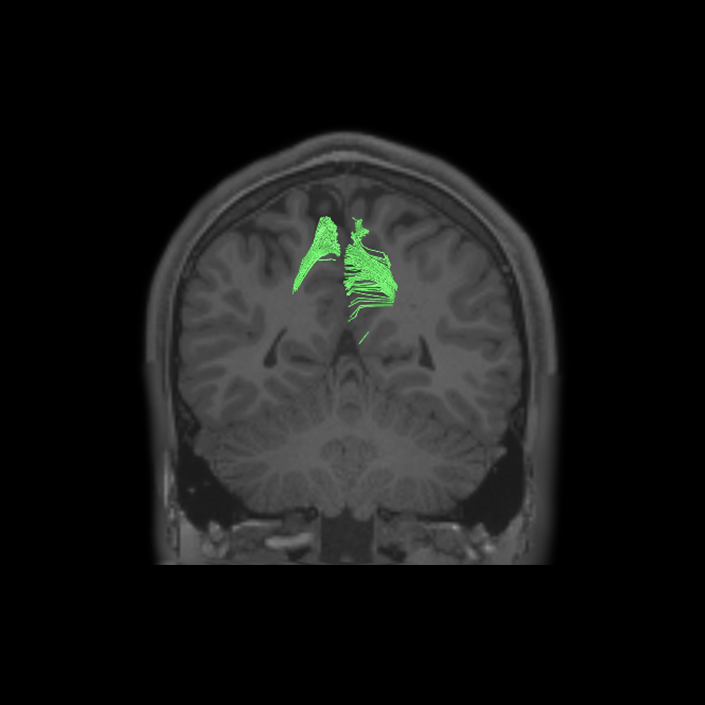
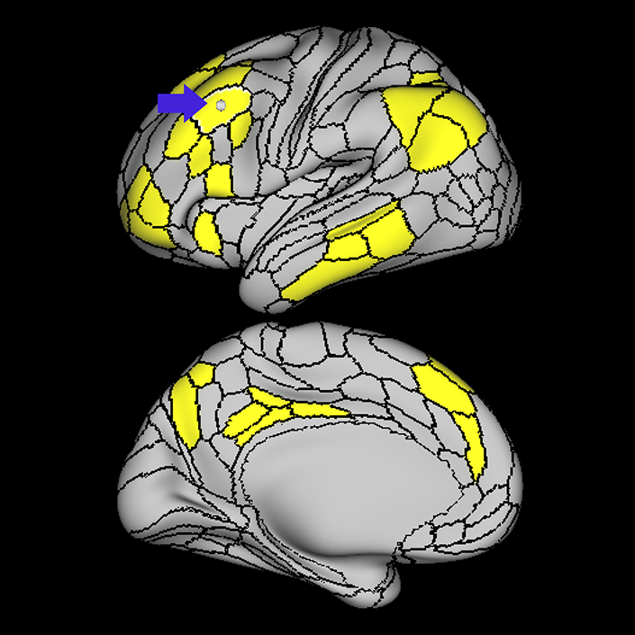
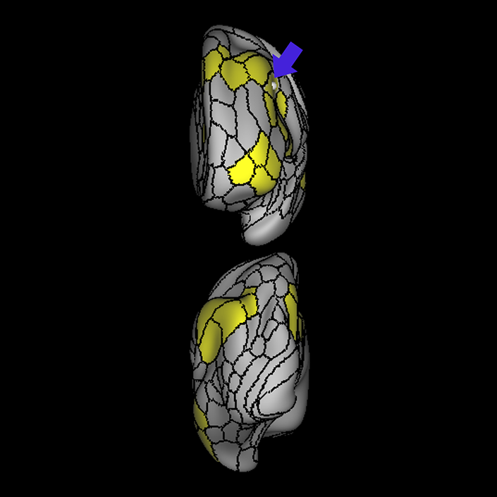
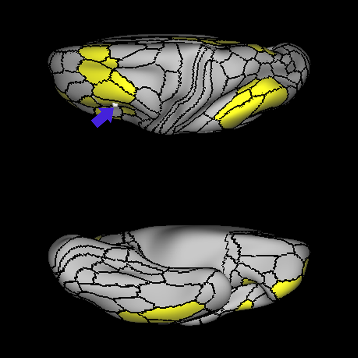
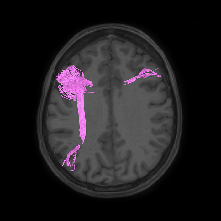
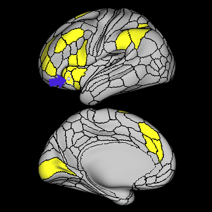
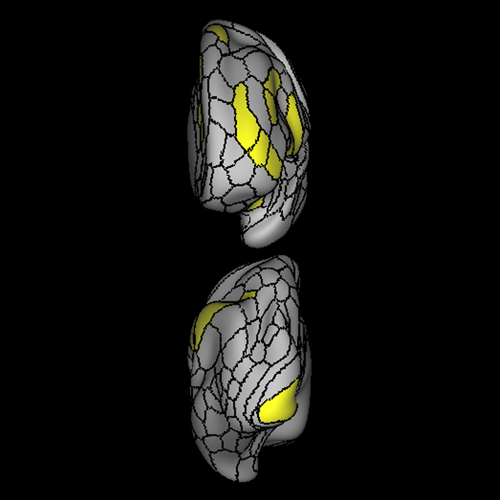
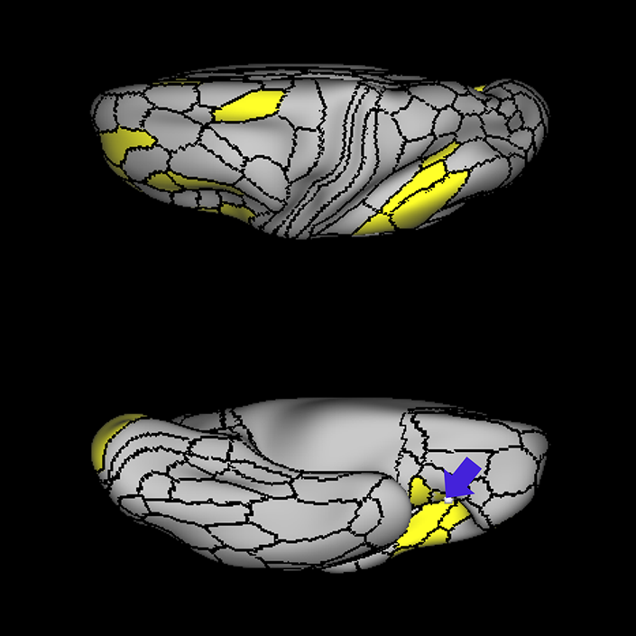
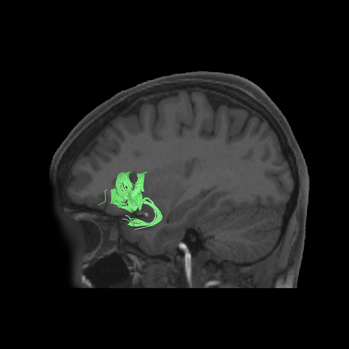
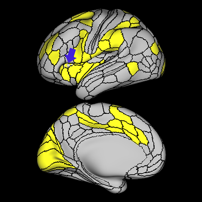
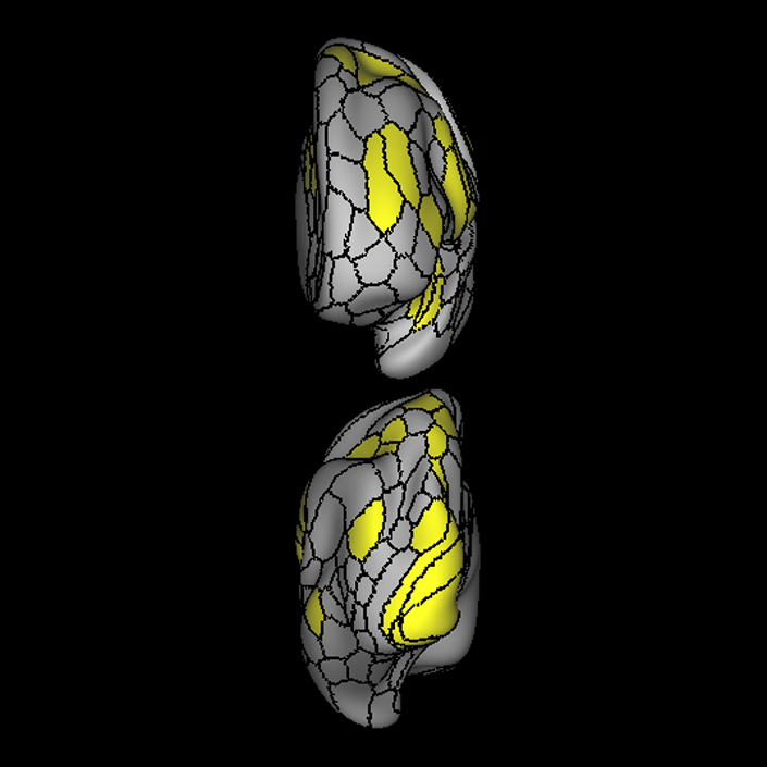
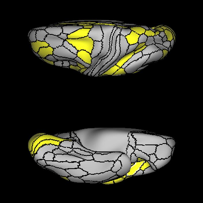
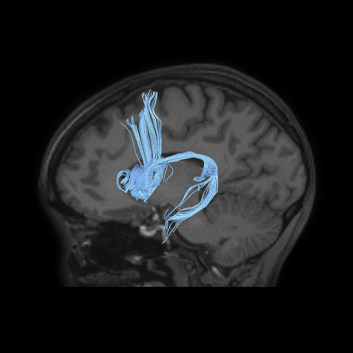
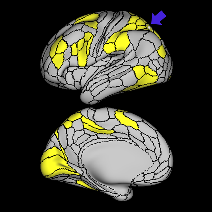
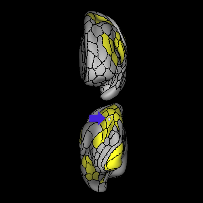
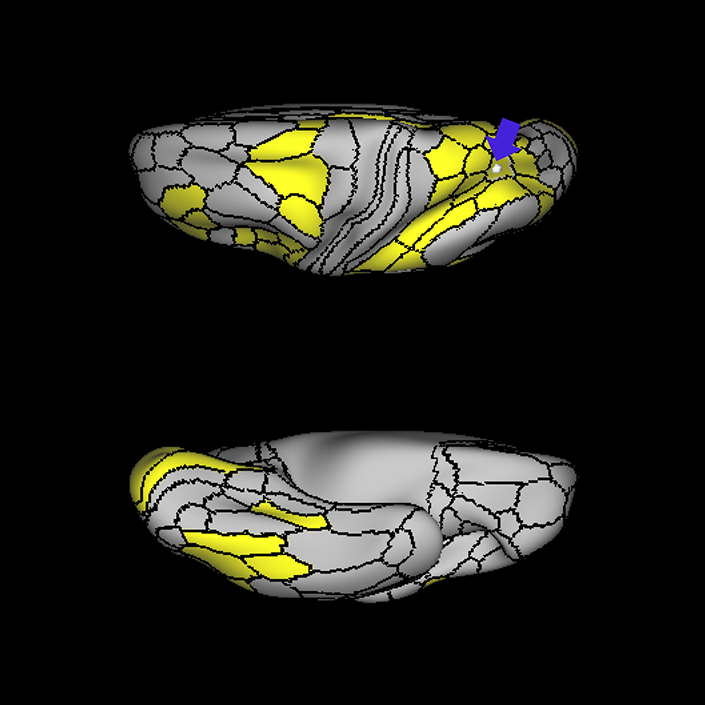
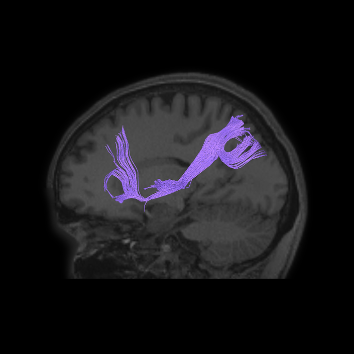
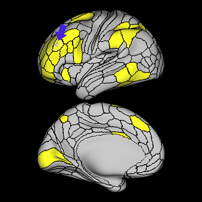
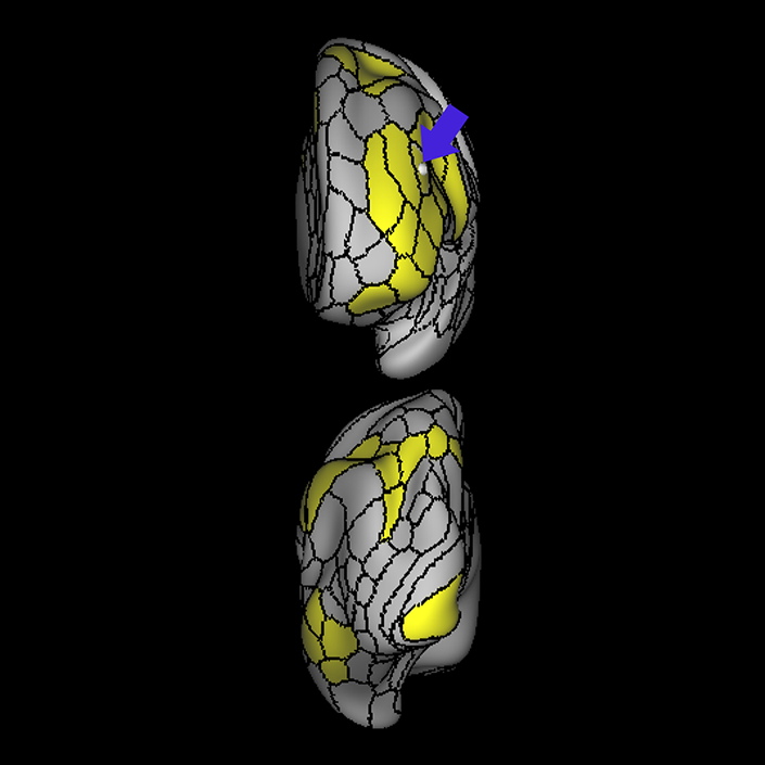
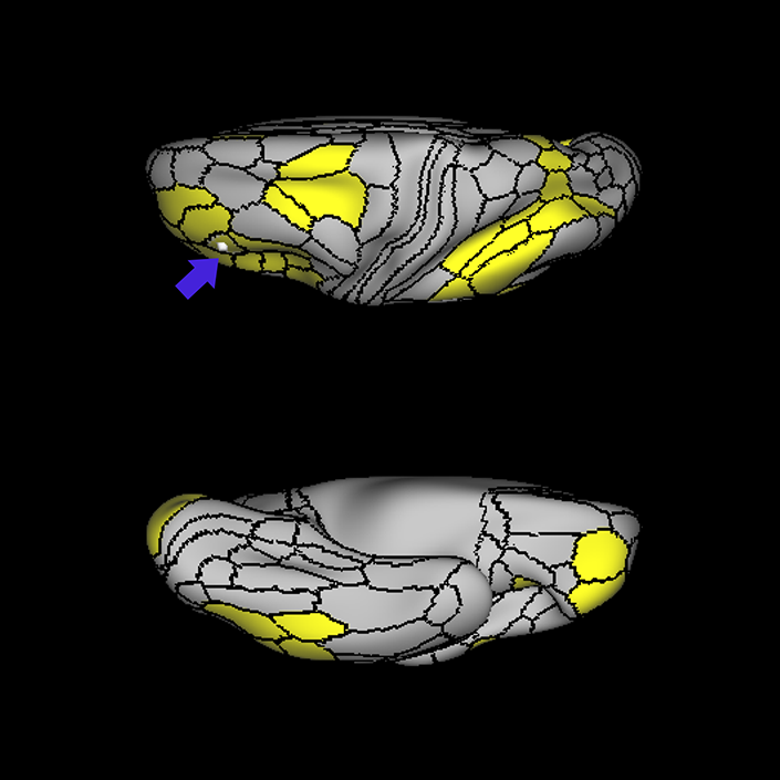
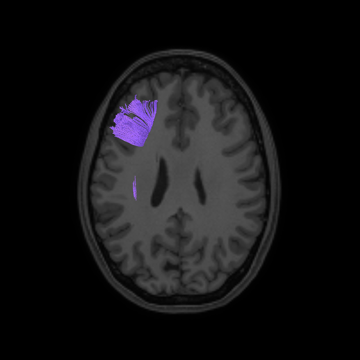
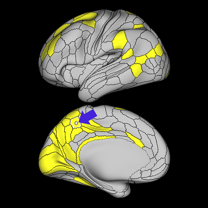
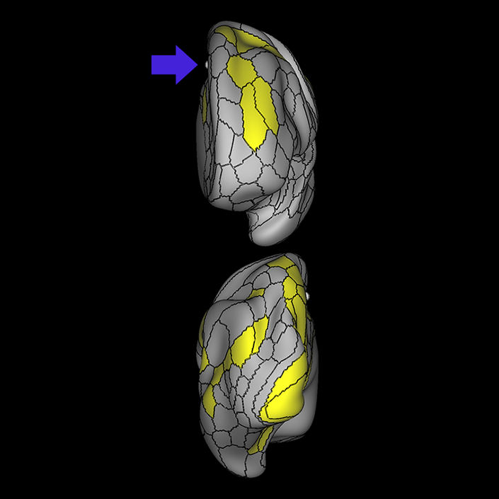
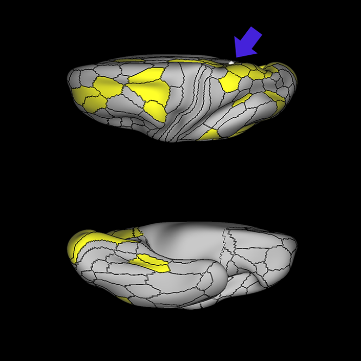
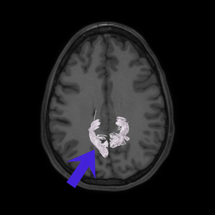
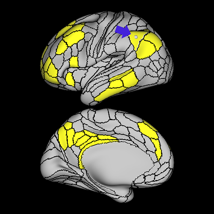
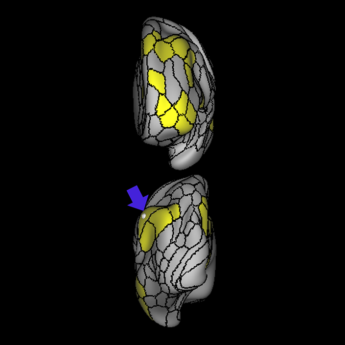
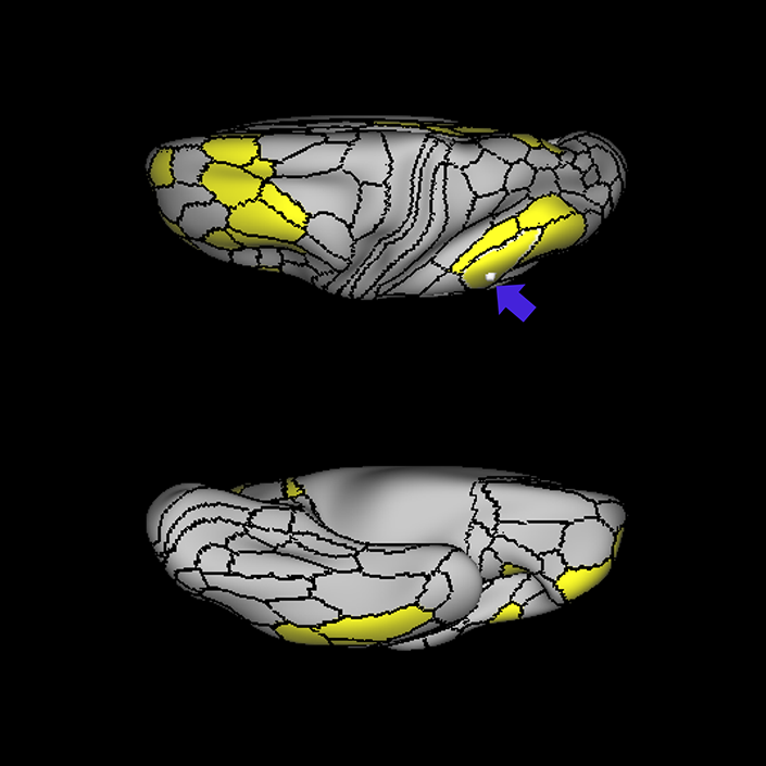
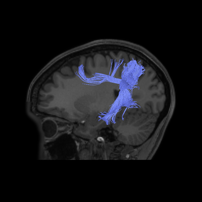
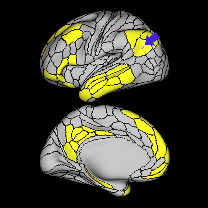
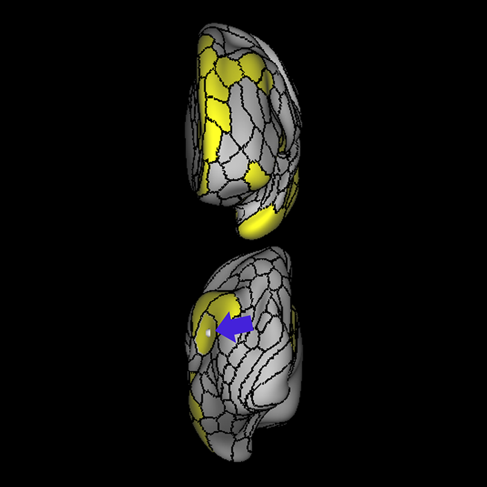
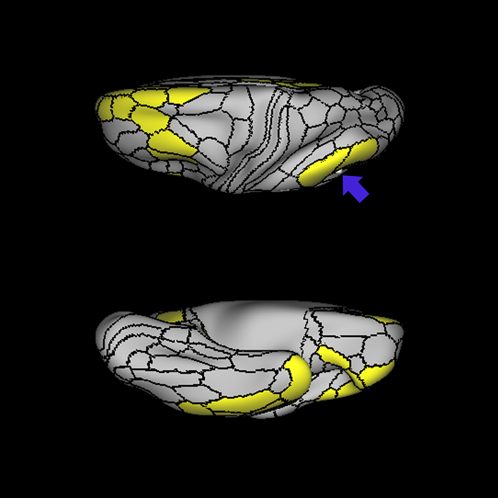
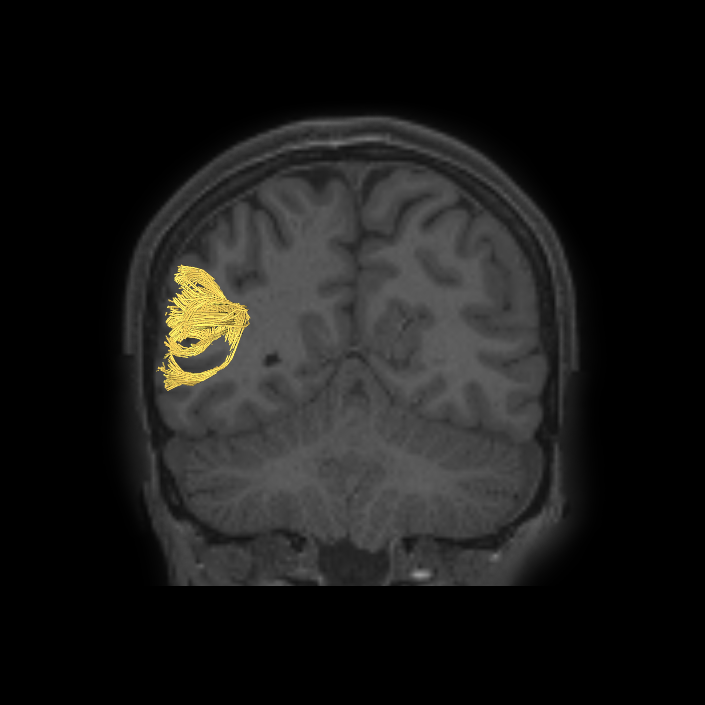
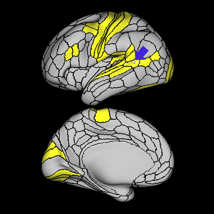
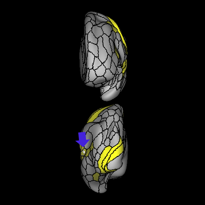
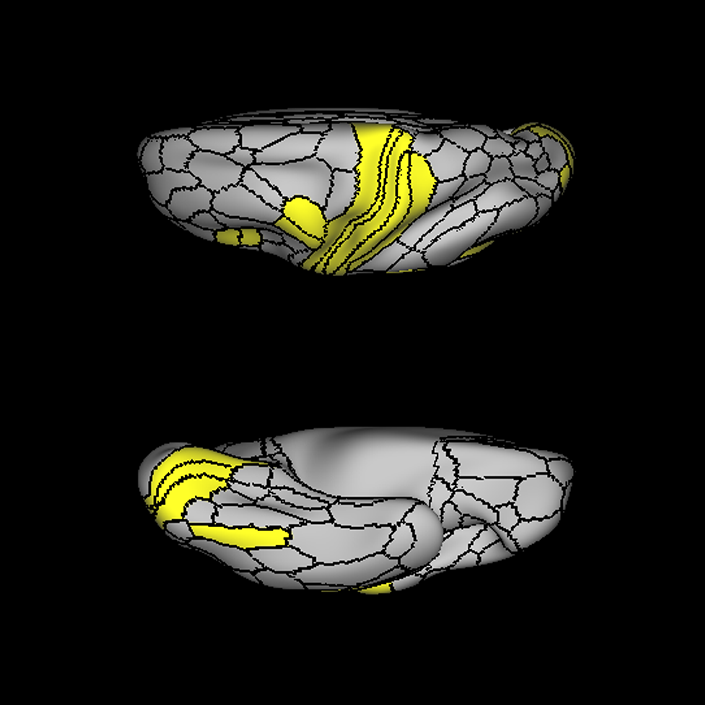
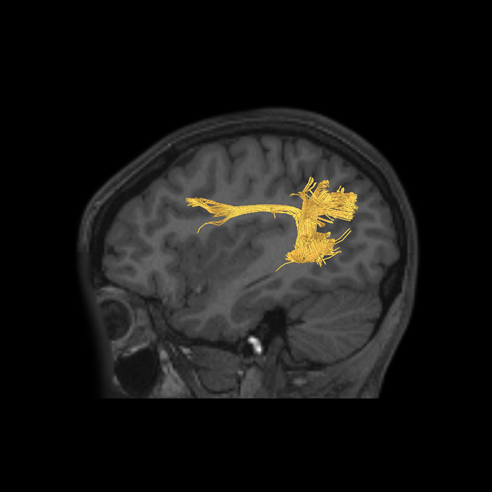
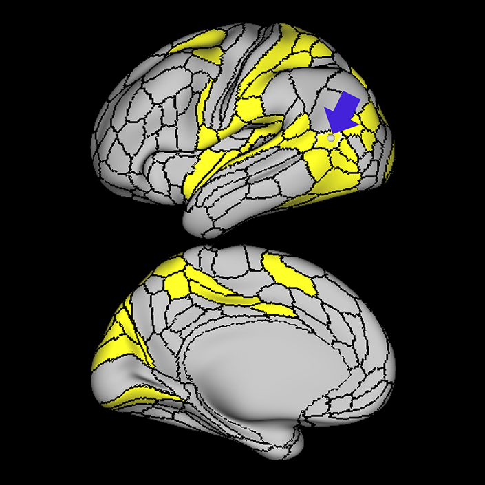
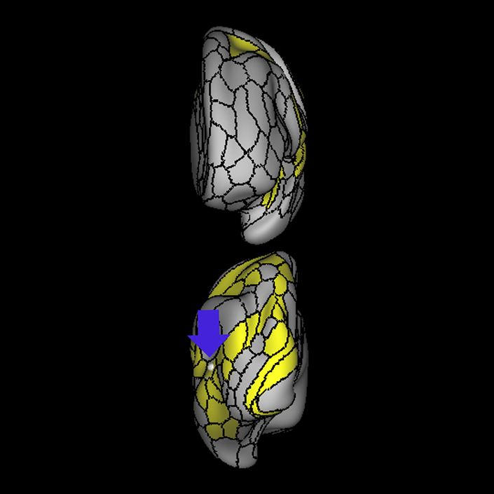
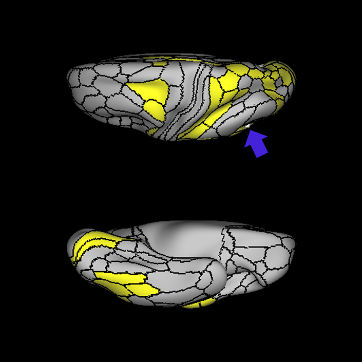
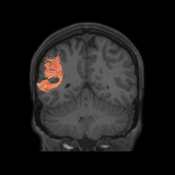
ᐅ SummaryArea 6a (6 anterior): part of the premotor areas. While the precise function of this area is unknown, the functions of the premotor cortex are well characterized in the literature. 6a shows greater activation when solving math problems, in social interaction settings, and when performing object feature comparison tasks. ᐅ Where is it?Area 6a (6 anterior) makes up the posterior superior most bank of the superior frontal sulcus and the adjacent portions of the superior frontal gyrus, principally forming this bank just as the sulcus forms the right angle with the precentral sulcus. ᐅ What are its borders?Area 6a borders areas 6d and 6mp posteriorly, and area 6ma medially. Its anterior border is made by s6-8, area 8AD, and i6-8. FEF is its inferior neighbor. ᐅ What are its functional connections?Area 6a demonstrates functional connectivity to area 2 in the sensory strip, areas SCEF, PEF, FEF, 6ma, 6mp, 6d, and 6v in the premotor regions, areas a24prime, p32prime, 5mv, and 23c in the middle cingulate regions, areas IFSa, IFJa, i6-8, 46, p9-46v and 9-46d in the lateral frontal lobe areas OP4, PFcm, FOP4, and FOP2 in the superior insula opercular regions, areas PoI1 and PoI2 in the lower opercula and Heschl's gyrus regions, areas TE2p, PHA3 and PHT in the temporal lobe, areas AIP, MIP, VIP, LIPd, LIPv, PFop, PF, PFt, PGp IP2, IP1, IP0, IPS1, 7AL,7PL, and 7PC, in the lateral parietal lobe, areas 7pm, 7am, DVT, and PCV in the medial parietal lobe, area V2 in the medial occipital lobe, and areas PH, TPOJ2, TPOJ3, and FST of the lateral occipital lobe. ᐅ What are its white matter connections?Area 6a is structurally connected to the pyramidal tracts and the parietal lobe. Connections to pyramidal tracts descend through the posterior limb of the internal capsule and cerebral peduncle to the brainstem. Parietal projections are portions of the SLF and connect with 3a, 3b, 7PC and 7AL. Local short association fibers connect with FEF, i6- 8, 55b, 8Av, 46 and 6r. ᐅ What is known about its function?Areas 6a and 6d are newly described subdivisions of the premotor cortex. While the precise function of these areas is unknown, the functions of the premotor cortex are well characterized in the literature. First, the premotor cortex is functionally divided into ventral and dorsal aspects. The dorsal premotor cortex is involved in associating informational cues with a particular body movement. These cues could be learned and arbitrary in nature or they can be based on other forms of somatosensation, such as visual or auditory sensation. The ventral premotor area is involved in hand movement manipulation of objects, e.g. when grasping or lifting. The ventral premotor area is also involved in more complex cortical functions such as when individuals learn actions or movements while observing others performing a task. Overall, the premotor cortex has significant function in the preparation of voluntary movements. Regarding area 6a more specifically, this region was distinguished from adjacent areas of the cortex based on differences in myelin thickness and functional activity.9 Compared to area s6-8, area 6a shows greater activation when solving math problems, in social interaction settings, and when performing object feature comparison tasks. Compared to area i6-8, area 6a shows greater activation in social interaction settings and relative deactivation in emotion identification and object feature comparison. Compared to area FEF, area 6a shows less activation in both gambling and object feature comparison. |
|
A: lateral-medial
B: anterior-posterior
C: superior-inferior
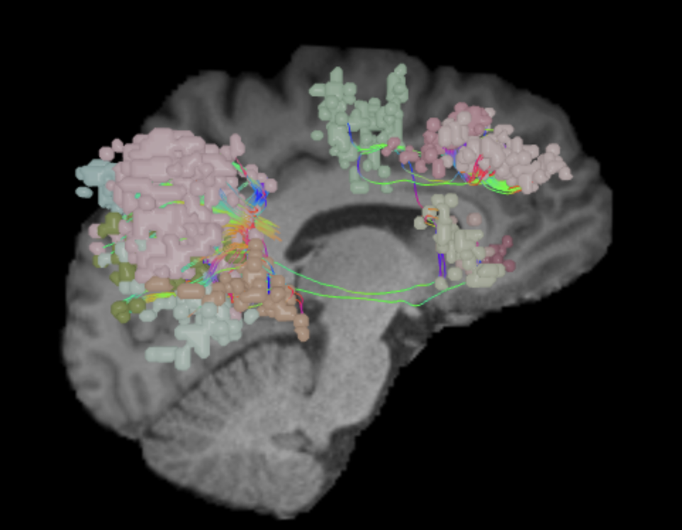
DTI image |
Parcellation Guide
Ventral Attention Network (VAN)
Why we think this network is worth considering in decision making:
Damage to the VAN results in hemispatial neglect, and other forms of cognitive dysfunction.
Evidence that this network is responsible for useful function in humans:
VAN encompasses lateral and inferior frontal/prefrontal cortex and the temporo-parietal junction (TPJ) and supporting stimulus driven attention.1 Spatial attention impairment is consistently associated with damage to the components of VAN.2
Consequences of damage to this network:
Hemispatial neglect: In the case of neglect, a close relation between ventral and dorsal system has been shown such that structural damage to the ventral attention areas are followed by functional impairments in the dorsal attention system.3 Studying stroke patients with damage to the components of VAN, He et al., (2007) provided evidence for a causal link between VAN and spatial neglect.4
Schizophrenia: Schizophrenia patients showed abnormal activation in the components of VAN.5
Depression: In a recent study, based on the relation between the effective connectivity measures and self-reported depression scores suggested VAN connectivity measures as a biomarker for depression in adolescents.6 Studies found reduced connectivity within the regions of VAN in children with a history of depression and/or anxiety.7
Eating disorders: Moreover, abnormal connectivity patterns between the VAN-DMN were found to be associated with binge-eating and purging behavior in adolescents with Blumia Nervosa (BN).8 Adult patients with BN and Anorexia Nervosa (AN) also displayed altered connectivity patterns in the VAN.9
Diabetes: VAN and DAN functional abnormalities are also related with the cognitive impairments associated with type 2 diabetes mellitus (T2DM),10 hepatic encephalopathy.11
Parkinson’s disease: In Parkinson’s Disease, in addition to the functional abnormalities in the attention network, minor hallucinations were associated with increased cortical thickness in VAN and DAN regions.12
ᐅ SummaryArea 6r (6 rostral): part of the premotor areas. Has not been extensively studied. The literature indicates that this area is functionally related to Broca's area in humans, which is a well-known cortical area essential to language processing. ᐅ Where is it?Area 6r (6 rostral) is the inferior portion of the precentral sulcus, including its floor and both banks. The latter point implies that some of area 6r lies on both the anterior inferior portion of the precentral gyrus and the posterior portion of the pars opercularis of the inferior frontal gyrus. The inferior extent of the precentral sulcus forms a shovel shaped cup near the opercular edge and this cup is area 6r. ᐅ What are its borders?Area 6r borders area 44 anteriorly and area 43 posteriorly. Area 6v forms a posterosuperior border with it. PEF is its main superior neighbor, while IFJp and IFJa form an anterior superior border. FOP1 and FOP4 form its inferior border on its undersurface opercular surface. ᐅ What are its functional connections?Area 6r demonstrates functional connectivity to areas SCEF, FEF PEF, 6ma, 6a and 6v in the premotor regions, areas 23c, 5mv, a24prime, p24prime,and p32prime in the middle cingulate regions, areas 46, p9-46v, 9-46d, IFSa, IFJa, IFJp, and p47r in the lateral frontal lobe, areas 43, OP4, PFcm, FOP2 FOP3, FOP4, and FOP5 in the superior insula opercular regions, areas AVI, MI, PoI1, and PoI2 in the lower opercula and Heschl's gyrus regions, areas TE2p and PHT in the temporal lobe, areas AIP, MIP, LIPd, LIPv, PFop, PF, PFt, IP0, 7PL, 7AL, and 7PC, in the lateral parietal lobe, and areas PH, and FST of the lateral occipital lobe. ᐅ What are its white matter connections?Area 6r is structurally connected to the superior longitudinal fasciculus and frontal aslant tract. FAT connects 6r with the superior frontal gyrus at parcellation SFL. Connections with the superior longitudinal fasciculus connect 6r to posterior temporal parcellations TE1a and TE2a. Local short association fibers connect with 6v, 44, IFJa, IFJp, 8C and IFSa. ᐅ What is known about its function?Area 6r has not been extensively studied. The literature indicates that this area is functionally related to Broca's area in humans, which is a well-known cortical area essential to language processing. |
|
A: lateral-medial
B: anterior-posterior
C: superior-inferior
DTI image |
ᐅ SummaryArea 7Am (7 anterior-medial): part of the lateral parietal lobe regions. Involved in several types of information processing including space, vision shape and motion, working memory, and execution. The anterior portion of 7Am is involved in self-centered mental imagery and attentional processes. Relative to its posterolateral neighbor 7PL, area 7Am is less activated during working memory and auditory story tasks. Relative to its posteromedial neighbor 7Pm, area 7Am is less activated during working memory and shape recognition tasks. ᐅ Where is it?Area 7AM (7 anterior-medial) is found on the anterosuperior surface of the medial face of the superior parietal lobule. ᐅ What are its borders?Area 7AM borders the area 7AL superior-laterally, VIP posterolaterally, and areas 7PL and 7PM posteriorly. On its interhemispheric face, its anterior border is made up of areas 5L, and 5MV. Its inferior border is made up of PCV (precuneus visual area), and its posterior border is made up of area 7PM. ᐅ What are its functional connections?Area 7AM demonstrates functional connectivity to areas SCEF, FEF, PEF, 6ma, 6a, and 6r in the premotor regions, areas IFSa, 46, and 9-46d areas in the lateral frontal lobe, areas a24prime, a32prime, p32prime, 5mv, and 23c in the medial frontal lobe, areas MI, PoI1, PoI2, PFcm and FOP4 in the insula opercular regions, areas PHA3, PHT, and TE2p, in the temporal lobe, areas 7PC, 7AL, 7PL, AIP, VIP, MIP, LIPd, PFop, PFt, PF, PGp, IP2, and IP0 in the lateral parietal lobe, areas 7pm, PCV, POS2, and DVT in the medial parietal lobe, area V1 in the medial occipital lobe, areas V6 in the dorsal visual stream areas, area FFC in the ventral visual stream areas, and areas PH, TPOJ2, TPOJ3, and FST in the lateral occipital lobe. ᐅ What are its white matter connections?Area 7AM is structurally connected to the contralateral hemisphere and thalamus. Some individuals have IFOF connections but these tracts are inconsistent. Contralateral connections course through the corpus callosum to end at 7AM and 7Pm. Thalamic connections project inferior through the posterolateral thalamus to the brainstem and superior colliculus. Local association bundles connect with VIP and 7PL. ᐅ What is known about its function?Area 7AM is involved in several types of information processing including space, vision shape and motion, working memory, and execution. The anterior portion of 7AM is involved in self-centered mental imagery and attentional processes. Relative to its posterolateral neighbor 7PL, area 7AM is less activated during working memory and auditory story tasks. Relative to its posteromedial neighbor 7PM, area 7AM is less activated during working memory and shape recognition tasks. |
|
A: lateral-medial
B: anterior-posterior
C: superior-inferior
DTI image |
ᐅ SummaryArea 7Pm (7 posterior-medial): part of the lateral parietal lobe regions. In the left hemisphere, this region is involved in vision motion, space, vision shape, attention, and working memory. In the right hemisphere, this region is involved in vision motion, space, vision shape, working memory, motor learning, execution, and attention. Area 7PM also plays a role in episodic memory retrieval and saccade-related activity. Relative to its superolateral neighbor 7PL, area 7PM is deactivated vs activated when viewing body images vs a compilation of tool, face and place images. Area 7PM is also less activated during emotional and social cue tasks compared to 7PL. ᐅ Where is it?Area 7PM (7 posterior-medial) occupies the posterior superior parietal lobule at the angle where the convexity surface of the SPL turns inferior to form its interhemispheric surface. 7PM occupies portions of both surfaces. ᐅ What are its borders?Area 7PM borders area 7AM anteriorly and area 7PL laterally on its superior surface. On its interhemispheric surface, it borders PCV anteroinferiorly, area 7M inferiorly, and POS2 (parieto-occipital sulcus 2) posteroinferiorly. ᐅ What are its functional connections?Area 7PM demonstrates functional connectivity to areas i6- 8, s6-8, 8AD, 8BM, 8C, a10p, p10p, 46, 9-46d, a9-46v, p9-46v, a32prime, 23c, and IFJp in the frontal lobe, areas 6a and 6ma in the premotor areas, areas PHA2, PreS, PHT and TE1p in the temporal lobe, areas PGp, PGs, PFm, IP0, IP1, IP2, AIP, MIP, LIPd, and 7PL in the lateral parietal lobe, and areas 7m, 7AM, PCV, DVT, 31a, POS1, POS2 and RSC in the medial parietal lobe. ᐅ What are its white matter connections?Area 7PM is structurally connected to the contralateral side and thalamus. Some individuals have IFOF connections but these are inconsistent. Contralateral connections course through the corpus callosum to end at parcellations 7Pm, POS2 and PCV. Thalamic connections project inferior through the posterolateral thalamus to the brainstem and superior colliculus. Local short association bundles connect with POS2, PCV and 7AM . ᐅ What is known about its function?The function of area 7PM in the left and right hemispheres is distinct. In the left hemisphere, this region is involved in vision motion, space, vision shape, attention, and working memory. In the right hemisphere, this region is involved in vision motion, space, vision shape, working memory, motor learning, execution, and attention. Area 7PM also plays a role in episodic memory retrieval and saccade-related activity. Relative to its superolateral neighbor 7PL, area 7PM is deactivated vs activated when viewing body images versus a compilation of tool, face and place images. Area 7PM is also less activated during emotional and social cue tasks compared to 7PL. |
|
A: lateral-medial
B: anterior-posterior
C: superior-inferior
DTI image |
ᐅ SummaryArea 8C: part of the lateral frontal lobe regions. Within the context of spatial working memory, area 8C is involved in the interpretation of complex visual information and attention. Areas 8 and rostral 6, as part of the posterior dorsolateral frontal areas, are also involved in the maintenance of spatial information. ᐅ Where is it?Area 8C is located at the posterior part of the middle frontal gyrus. It is an anterior- to-posterior band which is lateral to area 8AV. ᐅ What are its borders?Area 8C has area 8AV as its main medial border. Its lateral border is with 3 inferior frontal sulcus areas: IFSp, IFJa, and IFJp. Area 55b and PEF (precentral eyefield) are its posterior borders (and discussed in a different section). Area p9-46v and to a lesser extent area 46 are its anterior neighbors. ᐅ What are its functional connections?Area 8C demonstrates functional connectivity to areas s6-8, i6-8, a9-46v, p9-46v, a10p, 8BL, 8AD, and 8AV in the dorsolateral frontal lobe, areas 8BM and d32 in the medial frontal lobe, areas IFSp, IFJp, a47r, p47r, and 44 in the inferior frontal lobe, area AVI in the insula, areas TE1m, TE1p, TE2a, STSva, and STSvp in the temporal lobe, areas IP1, IP2, LIPd, PFm, PGi and PGs in the inferior parietal lobe, and areas 7pm, 31pv, 31a, POS2, 23d, and d23ab in the medial parietal lobe. ᐅ What are its white matter connections?Area 8C is structurally connected to the arcuate/SLF and the contralateral hemisphere. Contralateral connections travel through the corpus callosum to end at 8C. Connections with the arcuate/SLF project posteriorly and wrap around the sylvian fissure to the posterior temporal lobe to end at PH and PHT. ᐅ What is known about its function?Within the context of spatial working memory, area 8C is involved in the interpretation of complex visual information and attention. Areas 8 and rostral 6, as part of the posterior dorsolateral frontal areas, are also involved in the maintenance of spatial information. |
|
A: lateral-medial
B: anterior-posterior
C: superior-inferior
DTI image |
ᐅ SummaryArea AVI (anterior ventral insula): part of anterior apex regions. Suggested to have a role in sensation and control of autonomic nervous system processes as well as playing a role in human awareness, self-recognition, time perception, and perceptual decision making. ᐅ Where is it?Area AVI (anterior ventral insula) is located in anterior superior apex of the insula. ᐅ What are its borders?Area AVI borders area 47l anteriorly, areas 47s and AAIC inferiorly, MI posteriorly, and FOP4 and FOP5 superiorly. ᐅ What are its functional connections?Area AVI demonstrates functional connectivity to areas 6ma and 6r in the premotor regions, areas 44, p47r, 8C, a9-46v, p9-46v and 9-46d in the lateral frontal lobe, areas 8BM d32, and a32prime in the medial frontal lobe, areas FOP4, and FOP5 in the superior insula opercular regions, areas AAIC and MI in the lower opercula and Heschl's gyrus regions, areas TE2p, PHA3 and PHT in the temporal lobe, areas LIPd, PF, PFm, and IP2 in the lateral parietal lobe, area V1 in the medial occipital lobe. ᐅ What are its white matter connections?Area AVI is structurally connected to local parcellations. Local short association bundles connect to insular parcellations 47s, 47l, AAIC, FOP4, FOP5, and MI, and temporal pole parcellations TGd. ᐅ What is known about its function?Area AVI is a newly described area of the brain and was parcellated from the anterior insula. The anterior insula is suggested to have a role in sensation and control of autonomic nervous system processes as well as playing a role in human awareness, self-recognition, time perception, and perceptual decision making. Area AVI was parcellated from areas MI and AAIC in the anterior insula based on functional activity differences between regions related to motor, arithmetic, auditory language, and semantic tasks. |
|
A: lateral-medial
B: anterior-posterior
C: superior-inferior
DTI image |
ᐅ SummaryArea FOP4 (frontal operculum 4): part of anterior apex regions. The frontal operculum plays a key role in the initiation of language and lexical retrieval required for language learning. ᐅ Where is it?Area FOP4 (Frontal operculum 4) is located on the inner surface of the pars opercularis of the IFG. ᐅ What are its borders?Area FOP4 borders area 44 on its exterior surface as well as a small border with area 6r posterosuperiorly. Its posterior border is made up of FOP1 and FOP3. Its inferior border is with MI and AVI. Its anterior border is with FOP5. ᐅ What are its functional connections?Area FOP4 demonstrates functional connectivity to areas SCEF, FEF, PEF, 6v, 6a, 6ma, and 6r in the premotor regions, areas IFSa, 46, and 9-46d in the lateral frontal lobe, areas 5mv, 23c, 24dv, a24prime, p24prime, a32prime, and p32prime in the medial frontal lobe, areas 43, PFcm, OP4, FOP3, and FOP5 in the superior insula opercular regions, areas AVI, MI, PI, 52, PoI1, and PoI2 in the lower opercula and Heschl's gyrus regions, area PHT in the temporal lobe, areas 7AL, 7PL, AIP, LIPv, LIPd, PF, PFt, PGp, and PFop in the lateral parietal lobe, area V6 in the dorsal visual stream area, area 7am and DVT in the medial parietal lobe, areas V1, V2, and V3 in the medial occipital lobe. ᐅ What are its white matter connections?Area FOP4 is structurally connected to the frontal aslant tract and the arcuate/SLF. Connections from FOP4 with the frontal aslant tract project superior to the superior frontal gyrus to end at parcellations 6ma and SFL. Arcuate/SLF fibers project posteriorly above the insula, curving around the termination of the sylvain fissure to end at TGd and TE1a. From the arcuate/SLF there are also projections to the superior temporal gyrus that end at A4 and A5. Local short association fibers connect with AVI, FOP3, FOP5 and MI 14. White matter connections from FOP4 in the right hemisphere have less consistent connections with the arcuate/SLF ᐅ What is known about its function?Area FOP4 is a newly described area of the brain that was parcellated from the frontal operculum. The frontal operculum plays a key role in the initiation of language and lexical retrieval required for language learning. Area FOP4 was parcellated from areas AVI and FOP1 based on differences in functional activity in motor-based tasks. |
|
A: lateral-medial
B: anterior-posterior
C: superior-inferior
DTI image |
ᐅ SummaryArea MIP (medial intraparietal): part of the lateral parietal lobe regions. Visuomotor area presumed to be involved in the control of arm-reaching movements as well as prehension. Important for reception of proprioceptive signals. Area MIP neurons are modulated by reaching activity as well as visual and somatosensory stimulation. Important for the adjustment of reaching movements and correction of movement error. Important in the transformation of visual information into motor action for precise movement. ᐅ Where is it?Area MIP (Medial intraparietal) is found in the posterior portion of the superior bank of the intraparietal sulcus. It extends onto the superior surface of the adjacent portion of the superior parietal lobule. ᐅ What are its borders?Area MIP borders IPS1 posteriorly, area 7PL and VIP medially, IP1 and IP0 inferiorly, and LIPv and LIPd anteriorly. ᐅ What are its functional connections?Area MIP demonstrates functional connectivity to areas SCEF, FEF, PEF 6a, 6v, 6r, and 6ma in the premotor regions, areas IFSa, IFJa, IFJp, 46, and p9-46v in the lateral frontal lobe, areas 5mv, and 23c in the medial frontal lobe, areas PHA3, PHT and TE2p, in the temporal lobe, areas 7PC, 7PL, 7AL, AIP, VIP, LIPv, LIPd, PF, PFt, PGp, IP2, IP1, IP0, and IPS1 in the lateral parietal lobe, areas DVT, 7AM and 7pm in the medial parietal lobe, areas V1 and V2 in the medial occipital lobe, area FFC in the ventral visual stream areas, and areas PH and FST in the lateral occipital lobe. ᐅ What are its white matter connections?Area MIP is structurally connected to the IFOF, MdLF and local parcellations. IFOF connections travel from MIP through the posterior temporal lobe and extreme/external capsule to the frontal lobe to end at parcellations 8BL, 9p and 45. Connections with MdLF travel deep to the parietal lobe to the planum temporale to end at PBelt and MBelt. The majority of local short association bundles project inferior to 7PL, IP0, IP1 and IPS1. ᐅ What is known about its function?This visuomotor area is presumed to be involved in the control of arm-reaching movements as well as prehension. This region is important for reception of proprioceptive signals. Area MIP neurons are modulated by reaching activity as well as visual and somatosensory stimulation. Furthermore, this region is important for the adjustment of reaching movements and correction of movement error. This shows this region's importance in transformation of visual information into motor action for precise movement. |
|
A: lateral-medial
B: anterior-posterior
C: superior-inferior
DTI image |
ᐅ SummaryArea p9-46v (posterior 9-46 ventral): part of the lateral frontal lobe regions. Like area 46, plays a role in goal-directed higher- order cognitive processes. The mid-DLPFC, which includes areas 9-46 and 46, is also involved in the conscious, active control of planned behavior. ᐅ Where is it?Area p9-46v (posterior 9-46 ventral) is a small triangular shaped region located in the middle frontal gyrus. ᐅ What are its borders?Area p9-46v borders area 8C posteriorly. Its medial border is area 46. Its lateral border is made of IFSp and IFJa. ᐅ What are its functional connections?Area p9-46v demonstrates functional connectivity to area 46, 9-46d, a9-46v a10p, p10p, 6ma, i6-8, a47r, p47r, 8C, IFJp, IFJa, IFSp and IFSa in the dorsolateral frontal lobe, areas 8BM, and 33prime in the medial frontal lobe, areas 6a and 6r in the premotor area, area 11L in the orbitofrontal region, areas TE1p, TE2p, PH, and PHT in the temporal lobe, area AVI in the insula, areas LIPd, AIP, MIP, PFm, 7PL, IP0, IP1, and IP2 in the parietal lobe, area 7pm in the medial parietal lobe, and area V1 in the occipital lobe. ᐅ What are its white matter connections?Area p9-46v is structurally connected to the arcuate/SLF. Connections with the arcuate/SLF project posteriorly and wrap around the sylvian fissure to the inferior temporal gyrus to end at TE2a. Local short association bundles connect with 46, a9-46v, IFJa, IFSa, IFSp, 8C and 9-46d. ᐅ What is known about its function?Area 9-46d, like Area 46, plays a role in goal-directed higher-order cognitive processes. The mid-dorsolateral prefrontal cortex, which includes areas 9-46 and 46, is also involved in the conscious, active control of planned behavior. |
|
A: lateral-medial
B: anterior-posterior
C: superior-inferior
DTI image |
ᐅ SummaryArea PCV (precuneus visual area): part of the precuneus area. Involved in visual-spatial perception (including spatial reflection, visual motion perception, and spatial conflict resolution), episodic memory retrieval, self-processing, and consciousness. Involved in working memory processing of place, body, tool, and face images and recognizing emotional faces over neutral objects. ᐅ Where is it?Area PCV (Precuneus visual area) is found in the anterior precuneus, just posterior to the marginal ramus of the cingulate sulcus. ᐅ What are its borders?Area PCV borders areas 5mv, 23c and 31a anteriorly, area 31pd inferiorly, areas 7M and 7pm posteriorly, and area 7am superiorly. ᐅ What are its functional connections?Area PCV demonstrates functional connectivity to 46, 9-46d, 8AD, and in the lateral frontal lobe, areas a24prime, 5mv, 23c, and s32 in the medial frontal lobe, areas FEF, 6ma, and 6a in the premotor region, area STV in the insula opercular regions, areas PHA2, PHA3, and PHT in the temporal lobe, areas 7AL, 7PL, IP0, LIPd, PF, and PGp in the lateral parietal lobe, areas 23d, POS2, POS1, RSC, DVT, 7am, 7pm, 7m, 31a, and 31pd in the medial parietal lobe, areas V1, V2, in the medial occipital lobe, area V6 in the dorsal visual stream areas, and areas TPOJ2, TPOJ3 in the lateral occipital lobe. ᐅ What are its white matter connections?Area PCV is structurally connected to local parcellations and the contralateral hemisphere. Connections through the splenium of the corpus callosum terminate at contralateral 5m, 7am, PCV and 7AL. Short association bundles project superiorly to connect to 7am, 7pm and 5m. ᐅ What is known about its function?Area PCV is a part of the precuneus, which is involved in visual-spatial perception (including spatial reflection, visual motion perception, and spatial conflict resolution), episodic memory retrieval, self-processing, and consciousness. Task fMRI studies indicate that this region is specifically involved in working memory processing of place, body, tool, and face images and recognizing emotional faces over neutral objects. |
|
A: lateral-medial
B: anterior-posterior
C: superior-inferior
DTI image |
ᐅ SummaryArea PFm (parietal area F, part m): part of the lateral parietal lobe regions. Shows activation in nonspatial attention tasks, decision making when switching choices, rule change during visually guided attention, and reorientation. Also provide syntactical components to language processing, plays a role in attentional processing, and is activated in working memory, motor cue, and risk-related tasks. ᐅ Where is it?Area PFm (parietal area F, part m) is found on the anterior superior surface of the angular gyrus, and straddles the sulcus to lie on the posterior superior bank of the supramarginal gyrus. ᐅ What are its borders?Area PFm borders IP2 and IP1 superiorly, PF anteriorly, PSL and STV inferiorly, and PGI and PGS posteriorly. ᐅ What are its functional connections?Area PFm demonstrates functional connectivity to areas 8AV, 8AD, 8BL, 8C, s6-8, i6-8, a47r, p47r, a10p, p10p, 9a, a9-46v, p9-46v, and area 44 in the lateral frontal lobe, area d32 in the medial frontal lobe, area AVI in the insula, areas STSvp, TE1m, TE1p, and TE2a, in the temporal lobe, areas PGs, PGi, IP2, and IP1 in the lateral parietal lobe, and areas 7m, 7pm, POS2, 31a, 31pv, d23ab, 23d and RSC in the medial parietal lobe. ᐅ What are its white matter connections?Area PFm is structurally connected to the arcuate/SLF. Arcuate/SLF connections course anteriorly from PFm to 8C and 8BM, and inferiorly to middle temporal gyrus parcellations TE1a, TE1m, TE1p, STSva, STSvp and PHT. Local short association bundles connect with AIP, 7PC, IP1, IP2, LIPd, LIPv, PGi, PGs, 2 and 1 ᐅ What is known about its function?PFm shows activation in non-spatial attention tasks, decision making when switching choices, rule change during visually-guided attention, and reorientation. Intermediate regions of the inferior parietal lobule also provide syntactical components to language processing. This area also plays a role in attentional processing, and is activated in working memory, motor cue, and risk-related tasks. |
|
A: lateral-medial
B: anterior-posterior
C: superior-inferior
DTI image |
ᐅ SummaryArea PGi (parietal area G inferior): part of the lateral parietal lobe regions. Shows activity when individuals change their visuospatial attention from one area to another, and is a major node in the task-negative network, which mostly functions to redirect attention towards relevant stimuli. Relative to PGs, PGi is more active when processing faces compared to a body, is more active when listening to a story compared to unrelated words, and is more active when listening to a story vs answering arithmetic problems. ᐅ Where is it?Area PGi (parietal area G inferior) is found on the inferior surface of the angular gyrus. ᐅ What are its borders?Area PGi borders PGs superiorly, and PFm and STV anteriorly. Its inferior and its short posterior border is made up of TPOJ1, TPOJ2 and TPOJ3. ᐅ What are its functional connections?Area PGi demonstrates functional connectivity to areas 8AD, 8AV, 8BL, 8C, 47s, 47l, a47r, 44, 45, 10d, 9a, and 9p in the lateral frontal lobe, areas SFL, 9m, 10v, 10r, 8BM, and d32 in the medial frontal lobe, areas STSda, STSdp, STSva, STSvp, the hippocampus, TGd, TE1a, TE1m, TE1p, and TE2a, in the temporal lobe, areas PFm and PGs in the lateral parietal lobe, and areas 7m, POS1, 31a, 31pd, 31pv, d23ab, and v23ab in the medial parietal lobe. ᐅ What are its white matter connections?Area PGi is structurally connected to inferior parcellations of the occipitotemporal junction. Some individuals have connections to the arcuate/SLF with anterior projections to the premotor areas but this is inconsistent. Local short association bundles connect to the occipitotemporal junction at parcellations PHT, TE1p, STSvp, STSdp, TPOJ1 and TE1m. ᐅ What is known about its function?Area PGi is involved in many of the same functions as area PGs. This region shows activity when individuals change their visuospatial attention from one area to another, and is a major node in the task-negative network, which mostly functions to re-direct attention towards relevant stimuli. Relative to PGs, PGi is more active when processing faces compared to a body, is more active when listening to a story compared to un-related words, and is more active when listening to a story versus answering arithmetic problems. |
|
A: lateral-medial
B: anterior-posterior
C: superior-inferior
DTI image |
ᐅ SummaryArea TPOJ1 (temporo-parieto-occipital junction 1): part of the lateral parietal lobe regions. Participates in several processes including detecting incongruities between expected and presented stimuli, reward processing, and saliency. Also shows prominence in theory of mind, and the processing and detection of incongruous concepts and their subsequent resolution. Has specifically been shown to be activated during self-related processing, and receives input from different sensory afferent neurons. ᐅ Where is it?Area TPOJ1 (temporal-parietal-occipital junction 1) is found in the posterior superior temporal sulcus, just as it angles upward to indent the angular gyrus. It makes up both banks and the depth of this portion of the sulcus. ᐅ What are its borders?Area TPOJ1 borders STV superiorly, and PHT inferiorly. Its anterior border is small but borders STSdp, STSvp, A4, and A5. It borders PGi posterosuperiorly and TPOJ2 posteriorly. ᐅ What are its functional connections?Area TPOJ1 demonstrates functional connectivity to areas 1,2, 3a, and 3b in the sensory strip, area 4 in the motor strip, areas 55b and FEF in the premotor areas, areas IFJa and IFSp in the lateral frontal lobe, areas OP4, PFcm, RI, STV, PSL, A4, A5, PBelt, and LBelt in the insula opercular region, area STSdp in the temporal lobe, areas V2, V3, and V4 in the medial occipital lobe, area FFC in the ventral visual stream, and areas TPOJ2 and TPOJ3 in the lateral occipital lobe . ᐅ What are its white matter connections?Area TPOJ1 is structurally connected to the arcuate/SLF and inferior parietal lobe. Arcuate/SLF connections wrap around the sylvian fissure toward the fronal lobe to end at premotor parcellations IFJa, IFJp and 6r. From the arcuate/SLF there are also connections to the inferior parietal lobule that end at PFm, PGi and PGs. Local short association fibers connect with TPOJ2, STSvp and STSdp. ᐅ What is known about its function?The temporal-parietal-occipital junction participates in several processes including detecting incongruities between expected and presented stimuli, reward processing, and saliency. This region also shows prominence in theory of mind, and the processing and detection of incongruous concepts and their subsequent resolution. The TPOJ region has specifically been shown to be activated during self-related processing, and receives input from different sensory afferent neurons. The temporal-parietal-occipital junction is not finely parcellated in the current literature, thus task-based fMRI differences are the only way to delineate their individual functions. |
|
A: lateral-medial
B: anterior-posterior
C: superior-inferior
DTI image |
ᐅ SummaryArea TPOJ2 (temporo-parieto-occipital junction 2): part of the lateral parietal lobe regions. Shows greater activity when individuals are viewing an image of a body compared to an average image of places, tools, and faces. Is deactivated when viewing place images and when listening to a story vs unrelated words. ᐅ Where is it?Area TPOJ2 (temporal-parietal-occipital junction 2) is found on the anterior, superior part of the lateral occipital cortex. It straddles the sulcus and is on the posteroinferior bank of the angular gyrus. ᐅ What are its borders?Area TPOJ2 borders PGi superiorly, TPOJ1 anteriorly, PHT, FST and MST inferiorly, and TPOJ3 posteriorly. ᐅ What are its functional connections?Area TPOJ2 demonstrates functional connectivity to area 2 in the sensory strip, areas 6a, 6v, SCEF, and FEF in the premotor areas, areas 5mv, 23c and p24prime in the cingulate regions, areas OP4, 43, PFcm, 52, RI, STV, A4, PoI1, PoI2, PBelt in the insula opercular region, areas PHT and TE2p in the temporal lobe, areas PFop, PGp, IPS1, IP0, AIP, LIPv, 7AL, 7PL, and 7PC in the lateral parietal lobe, areas 7AM, PCV, and DVT in the medial parietal lobe, areas V2 and V3 in the medial occipital lobe, areas V3a, V6, and V6a in the dorsal visual stream, area FFC in the ventral visual stream, and areas FST, PH, LO3, MST, TPOJ1 and TPOJ3 in the lateral occipital lobe. ᐅ What are its white matter connections?Area TPOJ2 is structurally connected to the arcuate/SLF and inferior parietal lobule. Arcuate/SLF connections wrap around the sylvian fissure toward the fronal lobe to end at premotor parcellations IFJa, IFJp, 6v and 6r. From the arcuate/SLF there are also connections to the inferior parietal lobule that end at PFm, PGi and PGs. Local short association fibers connect with TPOJ1. ᐅ What is known about its function?Relative to its anterior neighbor TPOJ1, area TPOJ2 shows greater activity when individuals are viewing an image of a body compared to an average image of places, tools, and faces. TPOJ2 is deactivated when viewing place images and when listening to a story versus unrelated words. |
|
A: lateral-medial
B: anterior-posterior
C: superior-inferior
DTI image |
Reference list
- Asplund L, Todd J, Snyder P, Marois R. A central role for the lateral prefrontal cortex in goal-directed and stimulus-driven attention. Nature Neuroscience, 2010;13(4), 507–512. https://doi.org/10.1038/nn.2509
- Corbetta M, Shulman L. Spatial neglect and attention networks. Annual Review of Neuroscience, 2011;34, 569–599. https://doi.org/10.1146/annurev-neuro-061010-113731
- Corbetta M, Kincade J, Lewis C, Snyder Z, Sapir A. Neural basis and recovery of spatial attention deficits in spatial neglect. Nature Neuroscience, 2005;8(11), 1603–1610. https://doi.org/10.1038/nn1574
- He J, Snyder Z, Vincent L, Epstein A, Shulman L, Corbetta M. Breakdown of Functional Connectivity in Frontoparietal Networks Underlies Behavioral Deficits in Spatial Neglect. Neuron, 2007;53(6), 905–918. https://doi.org/10.1016/j.neuron.2007.02.013
- Jimenez M, Lee J, Wynn K, Cohen S, Engel A, Glahn C, Nuechterlein H, Reavis A, Green F. Abnormal ventral and dorsal attention network activity during single and dual target detection in schizophrenia. Frontiers in Psychology, 2016;7(MAR). https://doi.org/10.3389/fpsyg.2016.00323
- Liu J, Xu P, Zhang J, Jiang N, Li X, Luo Y. Ventral attention-network effective connectivity predicts individual differences in adolescent depression. Journal of Affective Disorders, 2019; 252, 55–59. https://doi.org/10.1016/j.jad.2019.04.033
- Sylvester M, Barch M, Corbetta M, Power D, Schlaggar L, Luby L. Resting state functional connectivity of the ventral attention network in children with a history of depression or anxiety. Journal of the American Academy of Child and Adolescent Psychiatry, 2013;52(12), 1326. https://doi.org/10.1016/j.jaac.2013.10.001
- Domakonda J, He X, Lee S, Cyr M, Marsh R. Increased Functional Connectivity Between Ventral Attention and Default Mode Networks in Adolescents With Bulimia Nervosa. Journal of the American Academy of Child and Adolescent Psychiatry, 2019;58(2), 232–241. https://doi.org/10.1016/j.jaac.2018.09.433
- Spalatro V, Amianto F, Huang Z, D’Agata F, Bergui M, Abbate Daga G, Fassino S, Northoff G. Neuronal variability of Resting State activity in Eating Disorders: increase and decoupling in Ventral Attention Network and relation with clinical symptoms. European Psychiatry, 2019;55, 10–17. https://doi.org/10.1016/j.eurpsy.2018.08.005
- Xia W, Wang S, Rao H, Spaeth M, Wang P, Yang Y, Huang R, Cai R, Sun H. Disrupted resting-state attentional networks in T2DM patients. Scientific Reports, 2015;5(1), 1–10. https://doi.org/10.1038/srep11148
- Chen F, Chen J, Liu J, Sun T, Shen T. Machine learning classification of cirrhotic patients with and without minimal hepatic encephalopathy based on regional homogeneity of intrinsic brain activity. PLoS ONE, 2016;11(3). https://doi.org/10.1371/journal.pone.0151263
- Sawczak M, Barnett J, Cohn M, Bologna M. Increased cortical thickness in attentional networks in Parkinson’s disease with minor hallucinations. Parkinson’s Disease, 2019. https://doi.org/10.1155/2019/5351749
