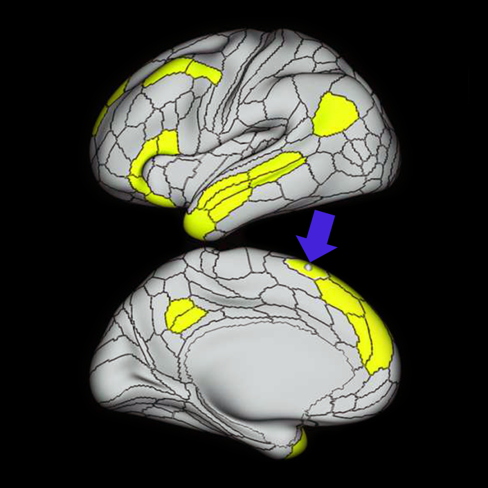
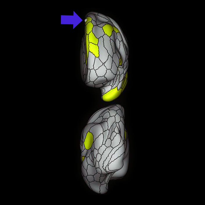
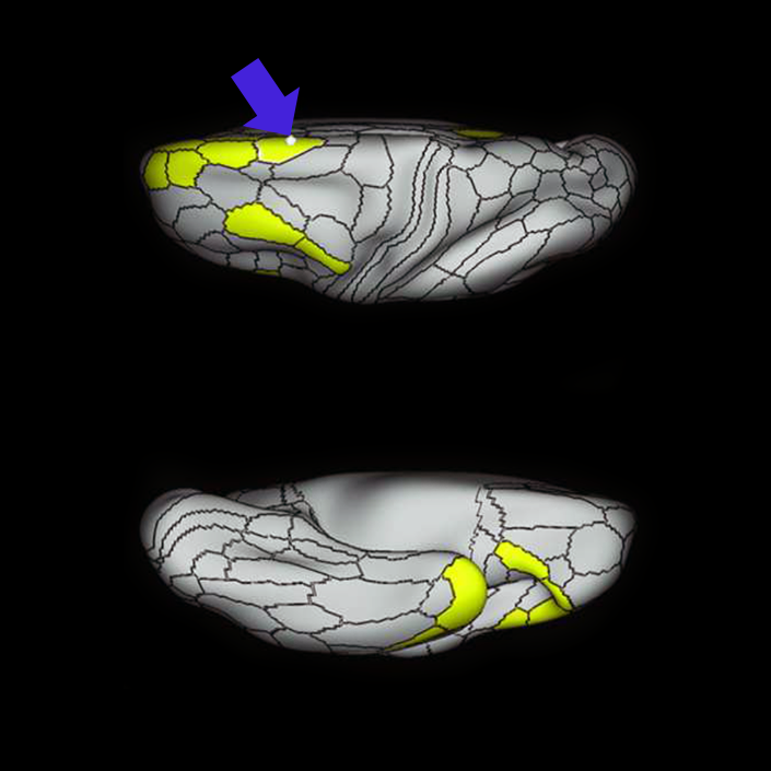
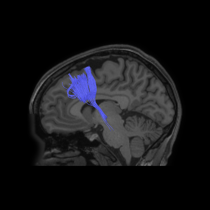
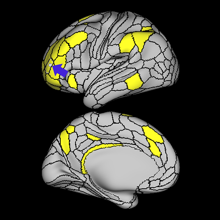
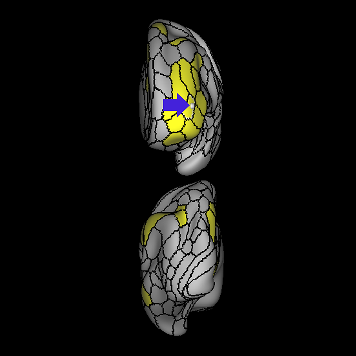
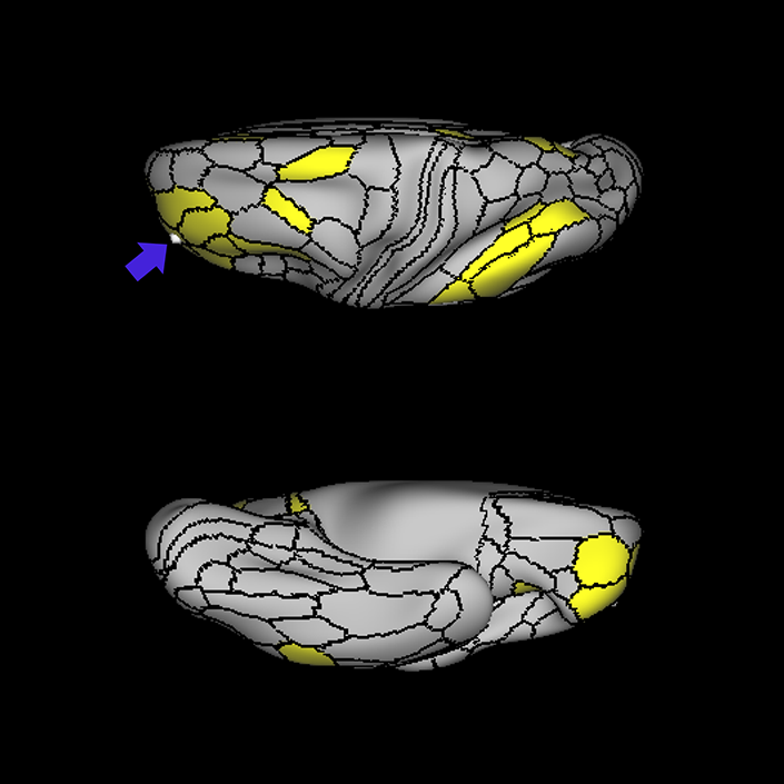
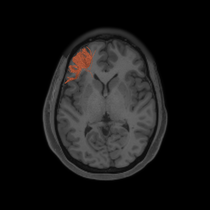
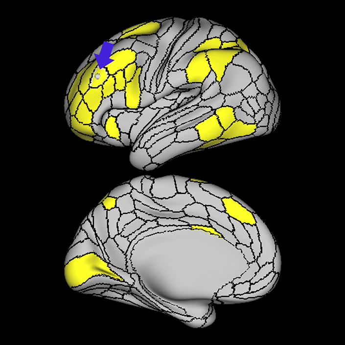
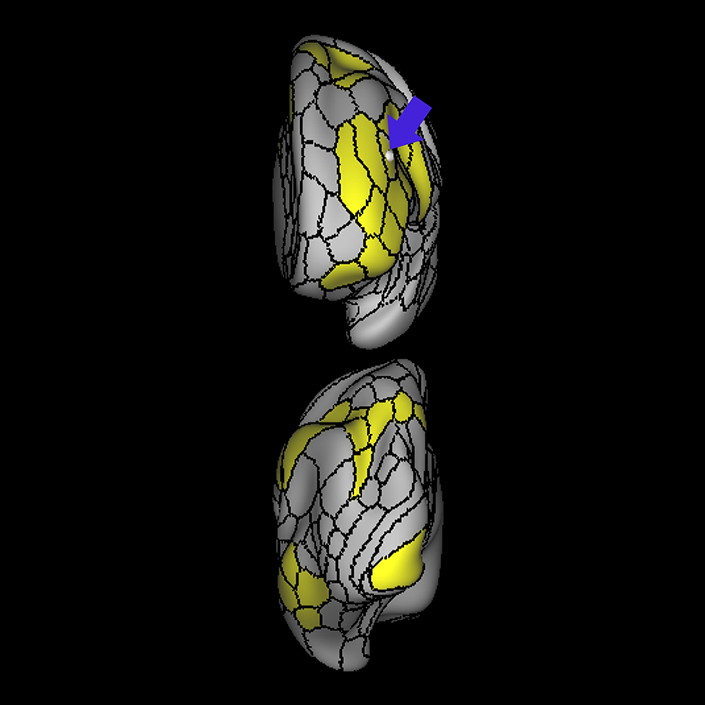
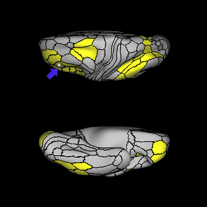
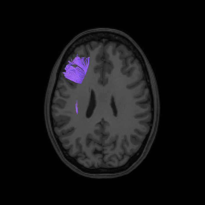
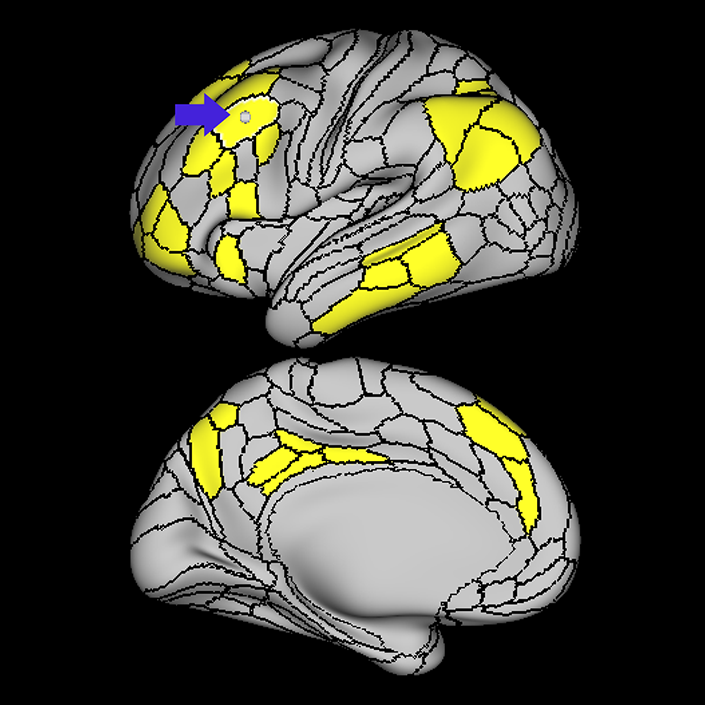
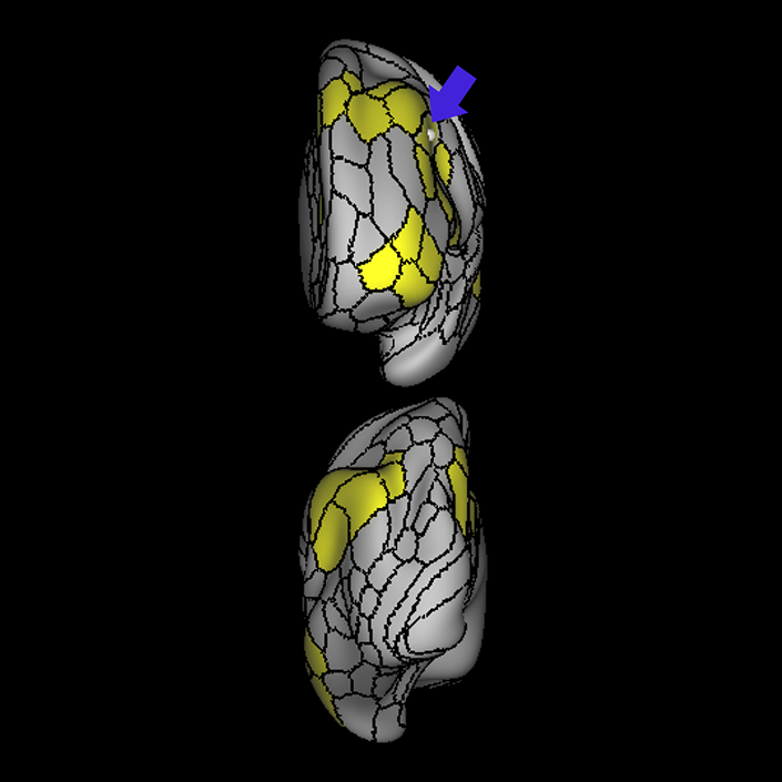
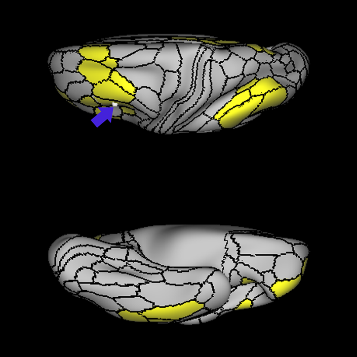
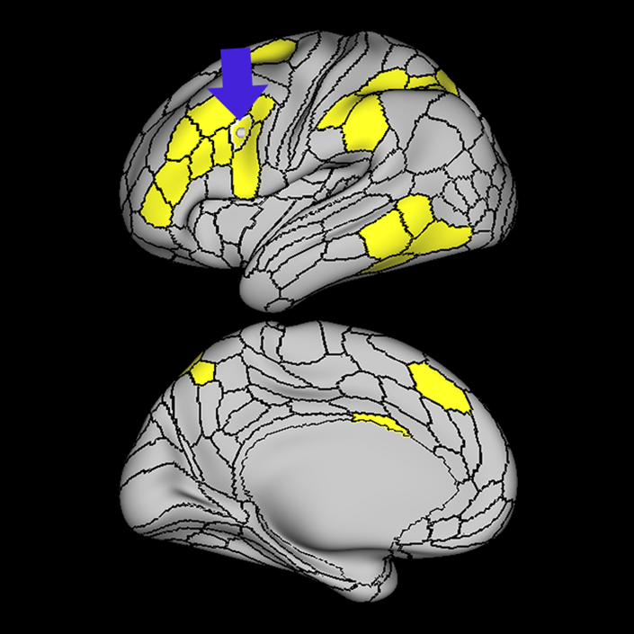
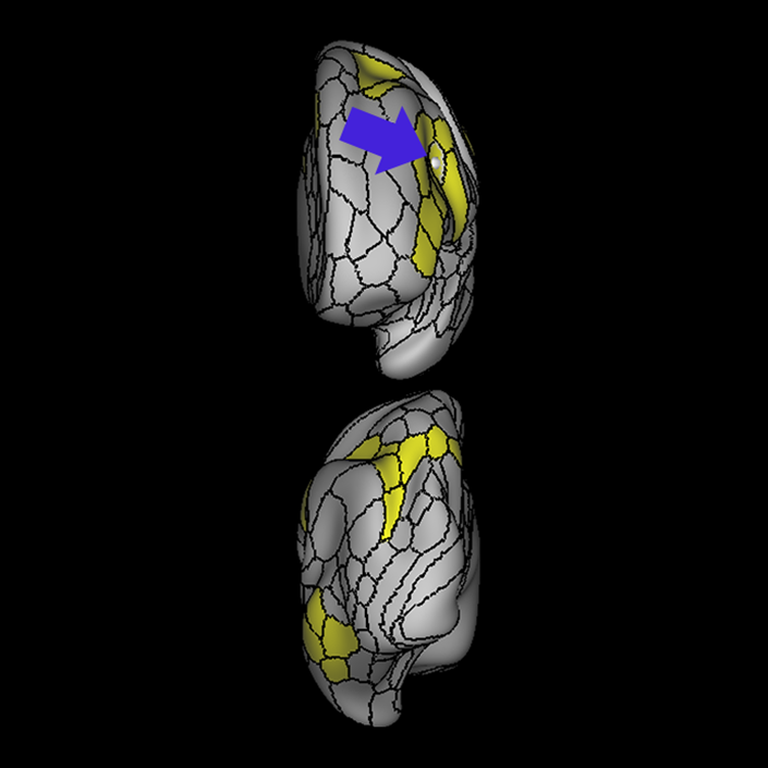
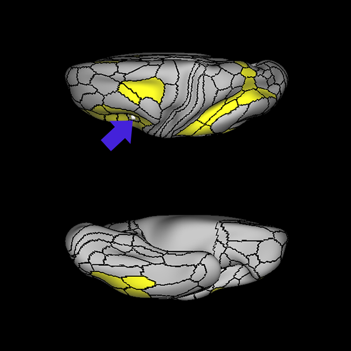
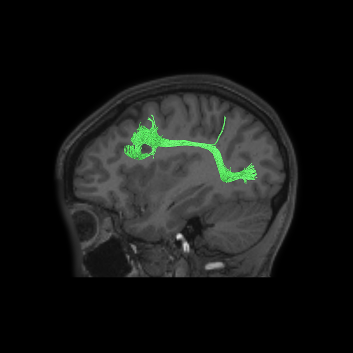
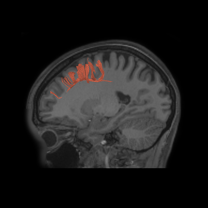
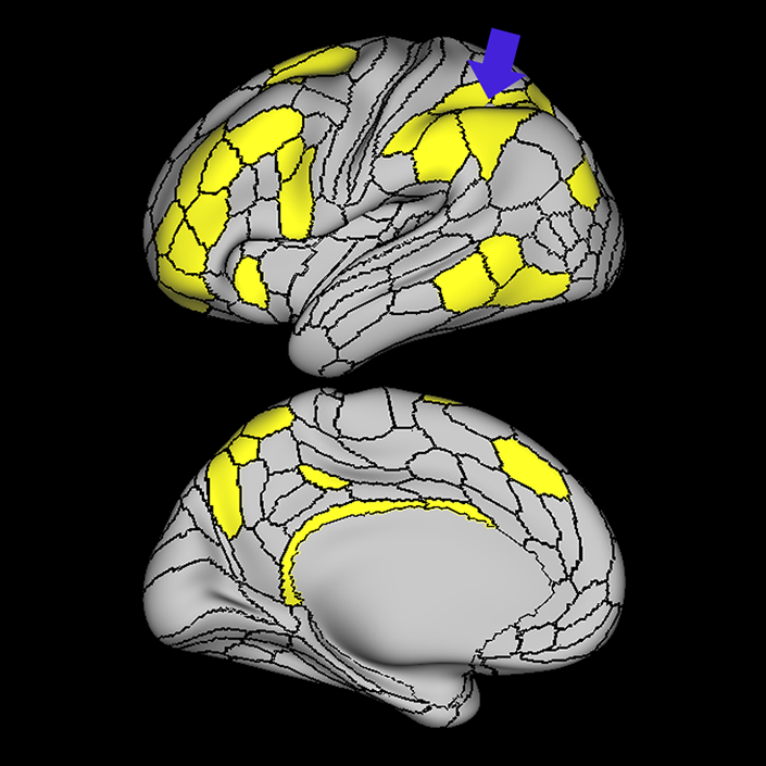
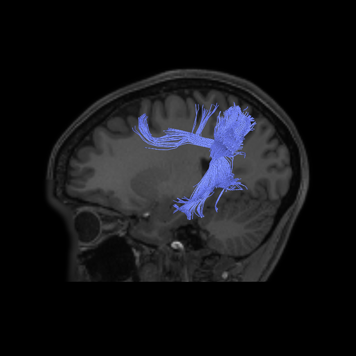
ᐅ SummaryArea a9-46v (anterior 9-46 ventral): part of the lateral frontal lobe regions. Like Area 46, plays a role in goal-directed higher- order cognitive processes. The mid- DLPFC, which includes areas 9-46 and 46, is also involved in the conscious, active control of planned behavior. ᐅ Where is it?Area a9-46v (anterior 9-46 ventral) is located at the anterior portior of the middle frontal gyrus. ᐅ What are its borders?Area a9-46v borders area 46 posteriorly, and area 9-46d medially. Its lateral border is mainly with area p47r and a slight contribution with IFSa. Its anterior limit forms a wedge with areas p10p and a47r. ᐅ What are its functional connections?Area a9-46v demonstrates functional connectivity to area 46, 9-46d, p9-46v, a10p, p10p, 6ma, i6-8, a47r, p47r, 8C, and IFSa in the dorsolateral frontal lobe, areas 8BM, and a32prime in the medial frontal lobe, area 11L in the orbitofrontal region, area TE1p in the temporal lobe, area AVI in the insula, areas LIPd, PF, IP1, and IP2 in the parietal lobe, and areas 7pm, 31a, and RSC in the medial parietal lobe . ᐅ What are its white matter connections?Area a9-46v is structurally connected to local parcellations. White matter tracts from this parcellation are highly variable. Short association bundles connect with 8C, 9-46d, 46, IFSa and p47r. ᐅ What is known about its function?Area 9-46d, like Area 46, plays a role in goal-directed higher-order cognitive processes. The mid-dorsolateral prefrontal cortex, which includes areas 9-46 and 46, is also involved in the conscious, active control of planned behavior. |
|
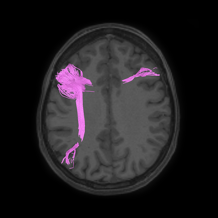
A: lateral-medial
B: anterior-posterior
C: superior-inferior
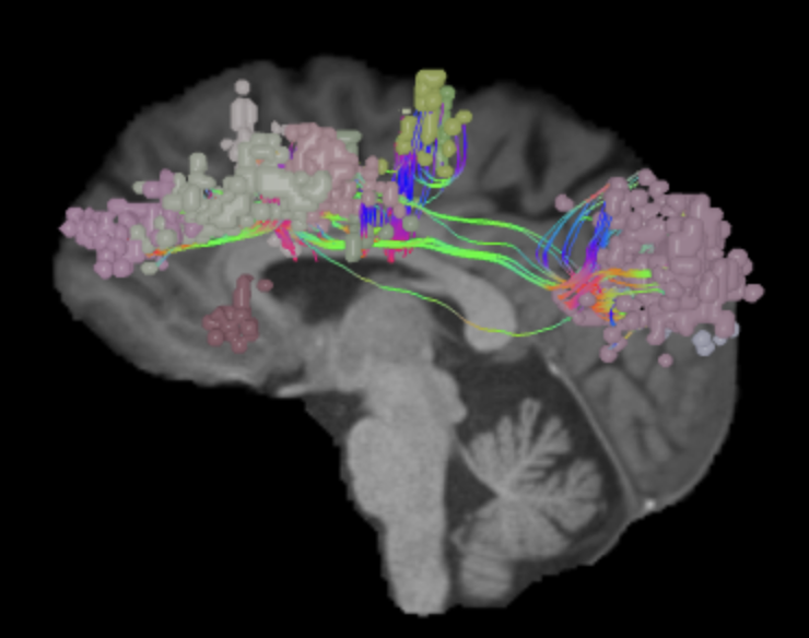
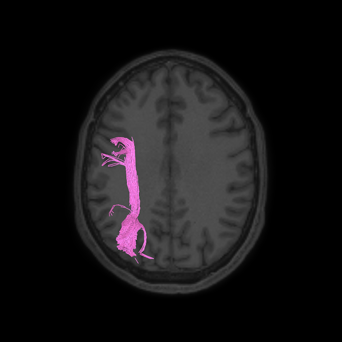
DTI image |
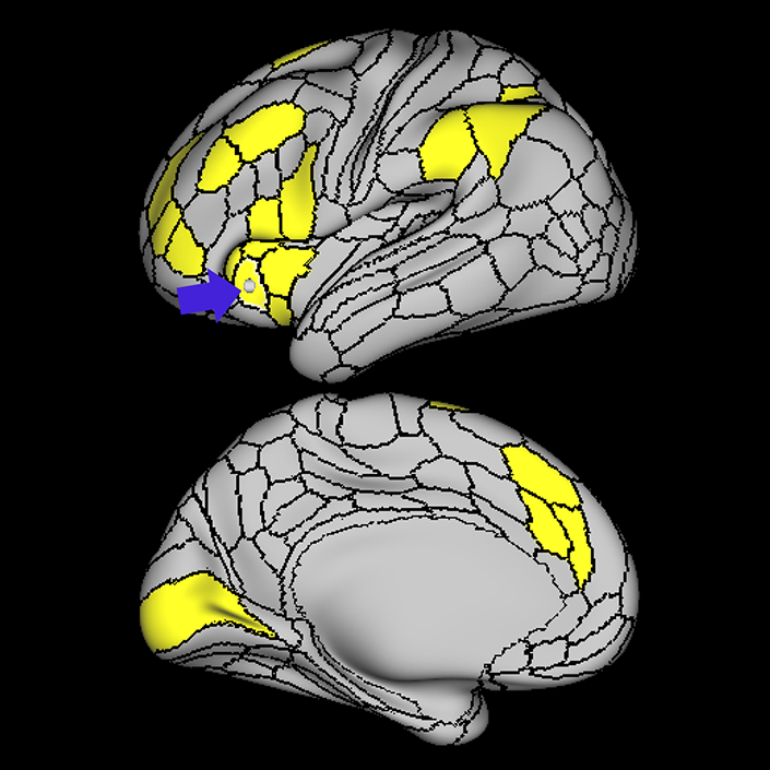
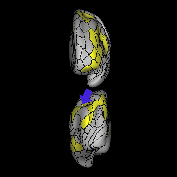
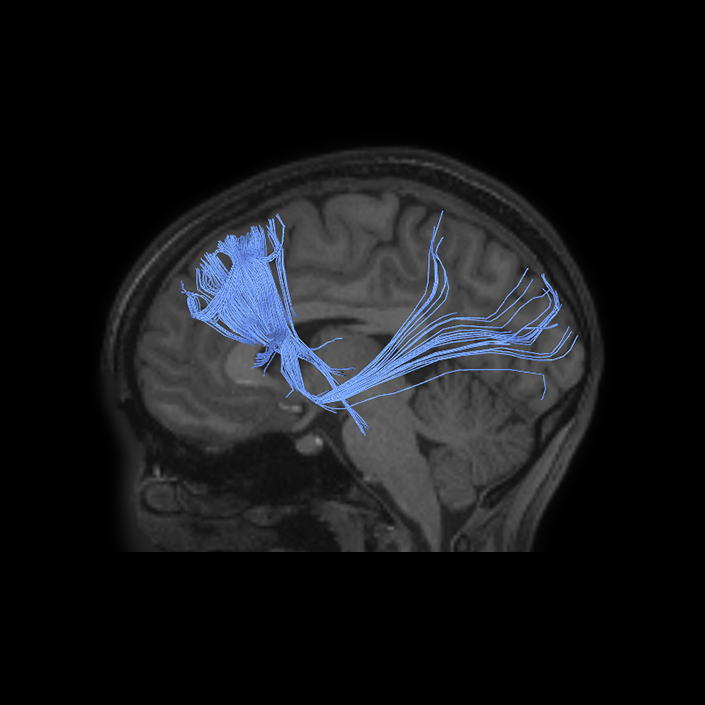
ᐅ SummaryArea p9-46v (posterior 9-46 ventral): part of the lateral frontal lobe regions. Like area 46, plays a role in goal-directed higher- order cognitive processes. The mid-DLPFC, which includes areas 9-46 and 46, is also involved in the conscious, active control of planned behavior. ᐅ Where is it?Area p9-46v (posterior 9-46 ventral) is a small triangular shaped region located in the middle frontal gyrus. ᐅ What are its borders?Area p9-46v borders area 8C posteriorly. Its medial border is area 46. Its lateral border is made of IFSp and IFJa. ᐅ What are its functional connections?Area p9-46v demonstrates functional connectivity to area 46, 9-46d, a9-46v a10p, p10p, 6ma, i6-8, a47r, p47r, 8C, IFJp, IFJa, IFSp and IFSa in the dorsolateral frontal lobe, areas 8BM, and 33prime in the medial frontal lobe, areas 6a and 6r in the premotor area, area 11L in the orbitofrontal region, areas TE1p, TE2p, PH, and PHT in the temporal lobe, area AVI in the insula, areas LIPd, AIP, MIP, PFm, 7PL, IP0, IP1, and IP2 in the parietal lobe, area 7pm in the medial parietal lobe, and area V1 in the occipital lobe. ᐅ What are its white matter connections?Area p9-46v is structurally connected to the arcuate/SLF. Connections with the arcuate/SLF project posteriorly and wrap around the sylvian fissure to the inferior temporal gyrus to end at TE2a. Local short association bundles connect with 46, a9-46v, IFJa, IFSa, IFSp, 8C and 9-46d. ᐅ What is known about its function?Area 9-46d, like Area 46, plays a role in goal-directed higher-order cognitive processes. The mid-dorsolateral prefrontal cortex, which includes areas 9-46 and 46, is also involved in the conscious, active control of planned behavior. |
|
A: lateral-medial
B: anterior-posterior
C: superior-inferior
DTI image |
ᐅ SummaryArea 8C: part of the lateral frontal lobe regions. Within the context of spatial working memory, area 8C is involved in the interpretation of complex visual information and attention. Areas 8 and rostral 6, as part of the posterior dorsolateral frontal areas, are also involved in the maintenance of spatial information. ᐅ Where is it?Area 8C is located at the posterior part of the middle frontal gyrus. It is an anterior- to-posterior band which is lateral to area 8AV. ᐅ What are its borders?Area 8C has area 8AV as its main medial border. Its lateral border is with 3 inferior frontal sulcus areas: IFSp, IFJa, and IFJp. Area 55b and PEF (precentral eyefield) are its posterior borders (and discussed in a different section). Area p9-46v and to a lesser extent area 46 are its anterior neighbors. ᐅ What are its functional connections?Area 8C demonstrates functional connectivity to areas s6-8, i6-8, a9-46v, p9-46v, a10p, 8BL, 8AD, and 8AV in the dorsolateral frontal lobe, areas 8BM and d32 in the medial frontal lobe, areas IFSp, IFJp, a47r, p47r, and 44 in the inferior frontal lobe, area AVI in the insula, areas TE1m, TE1p, TE2a, STSva, and STSvp in the temporal lobe, areas IP1, IP2, LIPd, PFm, PGi and PGs in the inferior parietal lobe, and areas 7pm, 31pv, 31a, POS2, 23d, and d23ab in the medial parietal lobe. ᐅ What are its white matter connections?Area 8C is structurally connected to the arcuate/SLF and the contralateral hemisphere. Contralateral connections travel through the corpus callosum to end at 8C. Connections with the arcuate/SLF project posteriorly and wrap around the sylvian fissure to the posterior temporal lobe to end at PH and PHT. ᐅ What is known about its function?Within the context of spatial working memory, area 8C is involved in the interpretation of complex visual information and attention. Areas 8 and rostral 6, as part of the posterior dorsolateral frontal areas, are also involved in the maintenance of spatial information. |
|
A: lateral-medial
B: anterior-posterior
C: superior-inferior
DTI image |
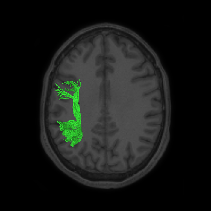
ᐅ SummaryArea IFJa (inferior frontal junction, posterior): part of the lateral frontal lobe. Areas in the midventrolateral prefrontal cortex interact with posterior areas of the brain to retrieve specific auditory memories. The inferior frontal junction, in particular, serves as an important crossroads between bottom-up and top-down processing in the lateral prefrontal cortex. ᐅ Where is it?Area IFJp is at the posterior most part of the inferior frontal sulcus. It is roughly superior to pars opercularis of the IFG. ᐅ What are its borders?Area IFJp borders IFJa anteriorly and the PEF posteriorly. Area 8C is its medial border and its inferior border is wedged between then upper borders of Areas 6R and 6V. ᐅ What are its functional connections?Area IFJp demonstrates functional connectivity to areas p47r, IFSa, IFJa, IFJp, p9-46v, i6-8, and 8C in the dorsolateral frontal lobe, areas 8BM and 33prime in the medial frontal lobe, areas 6r, 6a, and PEF in the premotor areas, areas PH, PHT, TE1p, and TE2p the temporal lobe, areas 7PL, PFt, PF, IP0, IP1, IP2, AIP, MIP and LIPd in the inferior parietal lobe, and area 7PM in the medial parietal lobe. ᐅ What are its white matter connections?Area IFJp is structurally connected with the arcuate/SLF and surrounding parcellations. Connections with the arcuate/SLF project posteriorly and wrap around the Sylvian fissure to the posterior temporal gyrus to end at PHT and FST. There are also connections from the arcuate/SLF to PFm. Local short association bundles connect with 8C, IFJa, IFJp, IFSp, 44, 6r and PEF. ᐅ What is known about its function?Areas in the midventrolateral PFC interact with posterior areas of the brain to retrieve specific auditory memories. The IFJ, in particular, serves as an important crossroads between bottom-up and top-down processing in the lateral prefrontal cortex. |
|
A: lateral-medial
B: anterior-posterior
C: superior-inferior
DTI image |
ᐅ SummaryArea i6-8 (inferior 6-8): part of the lateral frontal lobe. Areas s6-8 and i6-8 represent transitional areas of cortex between Brodmann areas 6 and 8. Areas 8 and rostral 6, as part of the posterior dorsolateral frontal areas, are involved in the maintenance of spatial information. Brodmann area 6 has also been subdivided into areas including the premotor and supplementary motor areas that influence motor control. The premotor area, which encompasses the lateral part of area 6, is further divided into ventral and dorsal portions, named PMv and PMd, respectively. ᐅ Where is it?Area i6-8 (inferior 6-8) is located at the posterior medial MFG near where the SFS joins with the precentral sulcus. ᐅ What are its borders?Area i6-8 borders area 6a medially, and area 55b and FEF laterally. Area 8AV is its lateral border. ᐅ What are its functional connections?Area i6-8 demonstrates functional connectivity to areas s6-8, a9-46v, p9-46v, p10p, 8C, 8AD, and 8AV in the dorsolateral frontal lobe, areas 8BM and d32 in the medial frontal lobe, areas IFSp, IFJp, a47r, p47r, and 44 in the inferior frontal lobe, area 6a in the premotor region, areas TE1m, TE1p, TE2a, PHA2, and PreS the temporal lobe, areas IP0, IP1, IP2, LIPd, PFm, and PGs in the inferior parietal lobe, and areas 7pm, 7m, 31a, POS2, POS1, and d23ab in the medial parietal lob. ᐅ What are its white matter connections?Area i6-8 is structurally connected to surrounding parcellations. White matter tracts from this parcellation are variable. Local short association bundles connect with 8Av, 8Ad, 9-46d, 6a and FEF. ᐅ What is known about its function?Areas s6-8 and i6-8 represent transitional areas of cortex between Brodmann Areas 6 and 8, previously described by Economo and Koskinas. Areas 8 and rostral 6, as part of the posterior dorsolateral frontal areas, are involved in the maintenance of spatial information. Brodmann Area 6 has also been subdivided into areas including the premotor and supplementary motor areas that influence motor control. The premotor area, which encompasses the lateral part of area 6, is further divided into ventral and dorsal portions, named PMv and PMd, respectively. |
|
A: lateral-medial
B: anterior-posterior
C: superior-inferior
DTI image |
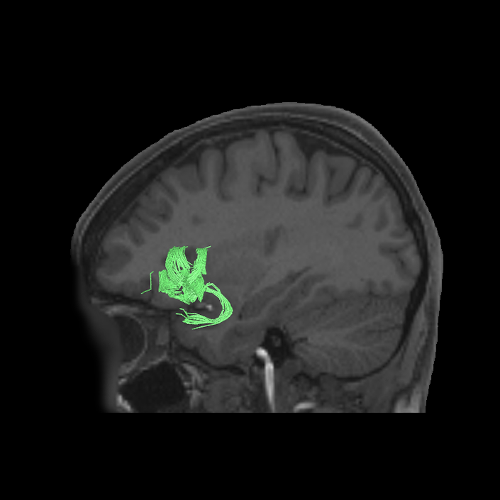
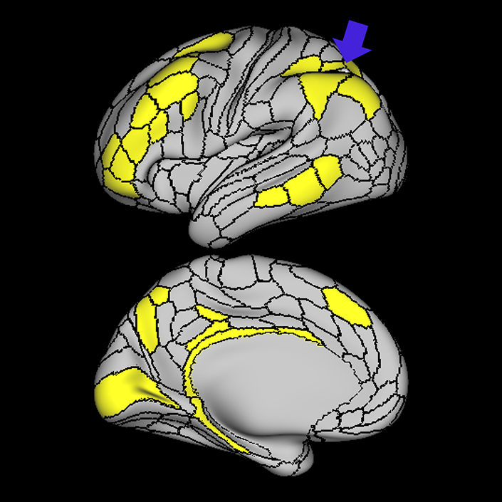
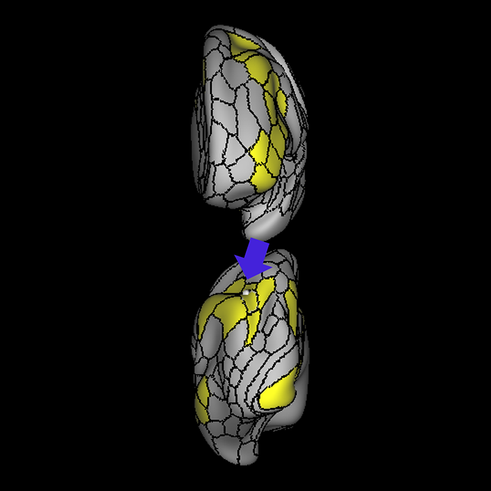
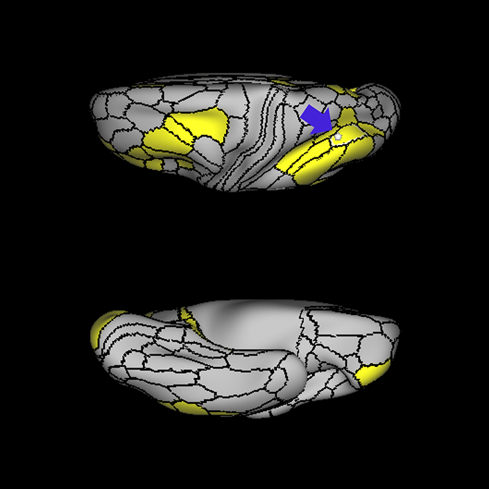
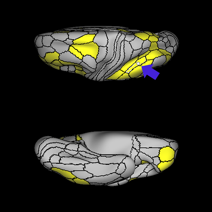
ᐅ SummaryArea AVI (anterior ventral insula): part of anterior apex regions. Suggested to have a role in sensation and control of autonomic nervous system processes as well as playing a role in human awareness, self-recognition, time perception, and perceptual decision making. ᐅ Where is it?Area AVI (anterior ventral insula) is located in anterior superior apex of the insula. ᐅ What are its borders?Area AVI borders area 47l anteriorly, areas 47s and AAIC inferiorly, MI posteriorly, and FOP4 and FOP5 superiorly. ᐅ What are its functional connections?Area AVI demonstrates functional connectivity to areas 6ma and 6r in the premotor regions, areas 44, p47r, 8C, a9-46v, p9-46v and 9-46d in the lateral frontal lobe, areas 8BM d32, and a32prime in the medial frontal lobe, areas FOP4, and FOP5 in the superior insula opercular regions, areas AAIC and MI in the lower opercula and Heschl's gyrus regions, areas TE2p, PHA3 and PHT in the temporal lobe, areas LIPd, PF, PFm, and IP2 in the lateral parietal lobe, area V1 in the medial occipital lobe. ᐅ What are its white matter connections?Area AVI is structurally connected to local parcellations. Local short association bundles connect to insular parcellations 47s, 47l, AAIC, FOP4, FOP5, and MI, and temporal pole parcellations TGd. ᐅ What is known about its function?Area AVI is a newly described area of the brain and was parcellated from the anterior insula. The anterior insula is suggested to have a role in sensation and control of autonomic nervous system processes as well as playing a role in human awareness, self-recognition, time perception, and perceptual decision making. Area AVI was parcellated from areas MI and AAIC in the anterior insula based on functional activity differences between regions related to motor, arithmetic, auditory language, and semantic tasks. |
|
A: lateral-medial
B: anterior-posterior
C: superior-inferior
DTI image |
ᐅ SummaryArea IP1 (intraparietal 1): part of the lateral parietal lobe regions. Shows significant activation during mental arithmetic activities. Supports more complex parts of numeric and mathematical information processing. Shows greater activation in primary contrasts, when interpreting motor cues and when hearing a story compared to when hearing arithmetic problems. More active when individuals are processing faces than when processing shapes, and is less deactivated when hearing a story vs unrelated words. ᐅ Where is it?Area IP1 (Intraparietal 1) is found on the middle portion of the inferior bank of the intraparietal sulcus. ᐅ What are its borders?Area IP1 borders IP2 anteriorly and IP0 posteriorly. Its inferior border is formed by PFm and PGs, and its superior border is MIP and LIPv. ᐅ What are its functional connections?Area IP1 demonstrates functional connectivity to area 6a in the premotor regions, areas 33prime and 8BM in the middle cingulate regions, areas IFSa, IFSp, IFJp, a9-46v, p9-46v, 8AV, 8C, i6-8, a47r, and p47r in the lateral frontal lobe, areas TE1m, TE1p, PHT, and PreS in the temporal lobe, areas TE1p and PHT in the temporal lobe, areas PFm, PGs, IP0, IP2, AIP, MIP, and LIPd in the lateral parietal lobe, areas 7pm, 31a, d23ab, POS2, and RSC in the medial parietal lobe, and area V1 in the occipital lobe. ᐅ What are its white matter connections?Area IP1 is structurally connected with the superior longitudinal fasciculus (SLF). Connections with the SLF project anteriorly to the premotor cortex to end at 55b, FEF and PEF. Local short association fibers connect with PFm, LIPd, IP0, IPS1and PGs. White matter tracts in the right hemisphere have more inferior connections with the inferior frontal gyrus. ᐅ What is known about its function?Area IP1 shows significant activation during mental arithmetic activities, and, as part of the anterior IPS, supports more complex parts of numeric and mathematical information processing. Area IP1 shows greater activation in primary contrasts, when interpreting motor cues and when hearing a story compared to when hearing arithmetic problems. Relative to area IP2, area IP1 is more active when individuals are processing faces than when processing shapes, and is less deactivated when hearing a story versus unrelated words. |
|
A: lateral-medial
B: anterior-posterior
C: superior-inferior
DTI image |
ᐅ SummaryArea 1P2 (intraparietal 2): part of the lateral parietal lobe regions. Shows significant activation during mental arithmetic activities. Appear to support more complex parts of numeric and mathematical information processing. Involved in the modulatory sensorimotor integration processes related to fine finger movements. ᐅ Where is it?Area IP2 (Intraparietal 2) is found on the anterior most portion of the inferior bank of the intraparietal sulcus. ᐅ What are its borders?IP2 borders PF anteriorly, PFm inferiorly, IP1 posteriorly, and areas AIP and LIPv superiorly. ᐅ What are its functional connections?Area IP2 demonstrates functional connectivity to areas 6ma, 6a, and 6r in the premotor regions, areas 33prime and 8BM in the middle cingulate regions, areas IFSa, IFJp, a9-46v, p9-46v, 46, 11L, 8C, i6-8, a47r, and p47r in the lateral frontal lobe, areas AVI in the insula regions, area PHT in the temporal lobe, areas TE1p and PHT in the temporal lobe, areas PFt, PF, PFm, PGp, IP0, IP1, AIP, MIP, LIPd, and 7PL in the lateral parietal lobe, areas 7AM, 7pm, 31a, POS2, and RSC in the medial parietal lobe, and area PH in the occipital lobe. ᐅ What are its white matter connections?Area IP2 is structurally connected with the superior longitudinal fasciculus (SLF). Connections with the SLF project anteriorly to the premotor cortex to end at 55b and PEF. Local short association fibers connect with PFm, LIPd, AIP and IP1. White matter tracts in the right hemisphere have more inferior connections with the inferior frontal gyrus. ᐅ What is known about its function?Area IP2 shows significant activation during mental arithmeti activities. Anterior IPS areas appear to support more complex parts of numeric and mathematical information processing. The anterolateral bank of the IPS is involved in the modulatory sensorimotor integration processes related to fine finger movements. |
|
A: lateral-medial
B: anterior-posterior
C: superior-inferior
DTI image |
ᐅ SummaryArea PFm (parietal area F, part m): part of the lateral parietal lobe regions. Shows activation in nonspatial attention tasks, decision making when switching choices, rule change during visually guided attention, and reorientation. Also provide syntactical components to language processing, plays a role in attentional processing, and is activated in working memory, motor cue, and risk-related tasks. ᐅ Where is it?Area PFm (parietal area F, part m) is found on the anterior superior surface of the angular gyrus, and straddles the sulcus to lie on the posterior superior bank of the supramarginal gyrus. ᐅ What are its borders?Area PFm borders IP2 and IP1 superiorly, PF anteriorly, PSL and STV inferiorly, and PGI and PGS posteriorly. ᐅ What are its functional connections?Area PFm demonstrates functional connectivity to areas 8AV, 8AD, 8BL, 8C, s6-8, i6-8, a47r, p47r, a10p, p10p, 9a, a9-46v, p9-46v, and area 44 in the lateral frontal lobe, area d32 in the medial frontal lobe, area AVI in the insula, areas STSvp, TE1m, TE1p, and TE2a, in the temporal lobe, areas PGs, PGi, IP2, and IP1 in the lateral parietal lobe, and areas 7m, 7pm, POS2, 31a, 31pv, d23ab, 23d and RSC in the medial parietal lobe. ᐅ What are its white matter connections?Area PFm is structurally connected to the arcuate/SLF. Arcuate/SLF connections course anteriorly from PFm to 8C and 8BM, and inferiorly to middle temporal gyrus parcellations TE1a, TE1m, TE1p, STSva, STSvp and PHT. Local short association bundles connect with AIP, 7PC, IP1, IP2, LIPd, LIPv, PGi, PGs, 2 and 1 ᐅ What is known about its function?PFm shows activation in non-spatial attention tasks, decision making when switching choices, rule change during visually-guided attention, and reorientation. Intermediate regions of the inferior parietal lobule also provide syntactical components to language processing. This area also plays a role in attentional processing, and is activated in working memory, motor cue, and risk-related tasks. |
|
A: lateral-medial
B: anterior-posterior
C: superior-inferior
DTI image |
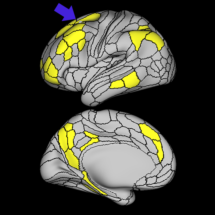
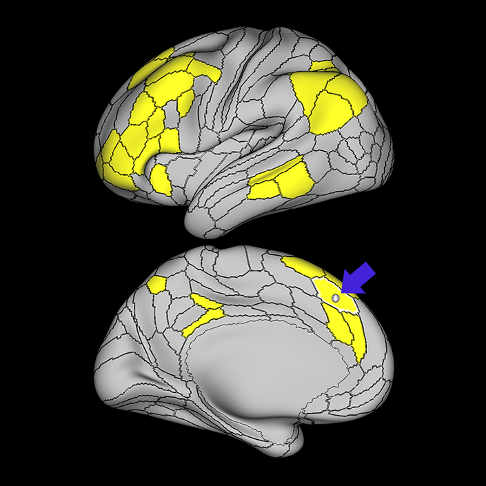
ᐅ SummaryArea 8BM (8B medial): part of medial superior frontal gyrus regions. Involved in maintaining visuospatial information as well as coordinating and coding visuospatial information in terms of oculomotor and other body-centered coordinate systems. ᐅ Where is it?Area 8BM (8B medial) is located in the posterior medial SFG. ᐅ What are its borders?Area 8BM borders area 9m anteriorly and SCEF posteriorly. It borders the following area 24 subdivisions superiorly: a24pr, p24pr, and p24. Its inferior border contains areas d32 and a32pr, and its superior boundary includes 8BL and SFL. ᐅ What are its functional connections?Area 8BM demonstrates functional connectivity to areas i6-8, s6-8, a10p, a9-46v, p9-46v, 8C, 8BL, 8AD, and 8AV in the dorsolateral frontal lobe, areas SFL, a32prime and d32 in the medial frontal lobe, areas IFSa, IFSp, IFJp, 44, 45, a47r, and p47r in the inferior frontal lobe, area 55b in the premotor areas, area AVI in the insula, areas TE1m, TE1p, and STSvp in the temporal lobe, areas LIPv, IP1, IP2, PFm, PGi and PGs in the lateral parietal lobe, and areas 7pm, 31a, and d23ab in the medial parietal lobe . ᐅ What are its white matter connections?Area 8BM is connected to the contralateral hemisphere, frontal aslant tract, inferior front-occipital fasciculus and thalamus. Contralateral connections course through the corpus callosum to end at 8BM and 9m. Frontal aslant tract fibers from 8BM project inferolaterally to end at 44. Thalamic connections run inferior to 8BM and continue in the brainstem. Fibers with the inferior fronto-occipital fasciculus project inferior and posterior through the extreme/external capsule through the temporal lobe to end at parietal and occipital connections 7PC, V1, V2 and V3. Local short association bundles connect with 9m, d32 and SCEF. ᐅ What is known about its function?Area 8Bm is involved in maintaining visuospatial information as well as coordinating and coding visuospatial information in terms of oculomotor and other body-centered coordinate systems. |
|
A: lateral-medial
B: anterior-posterior
C: superior-inferior
DTI image |
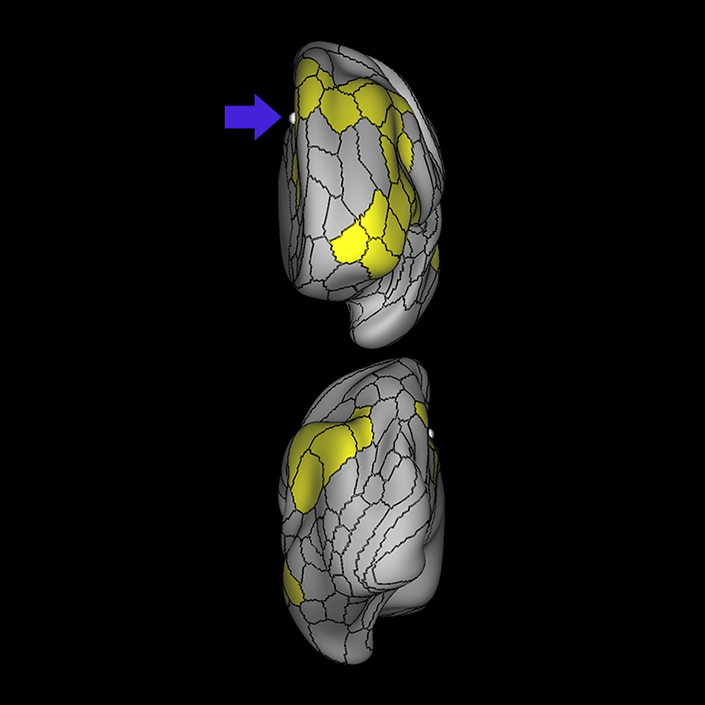
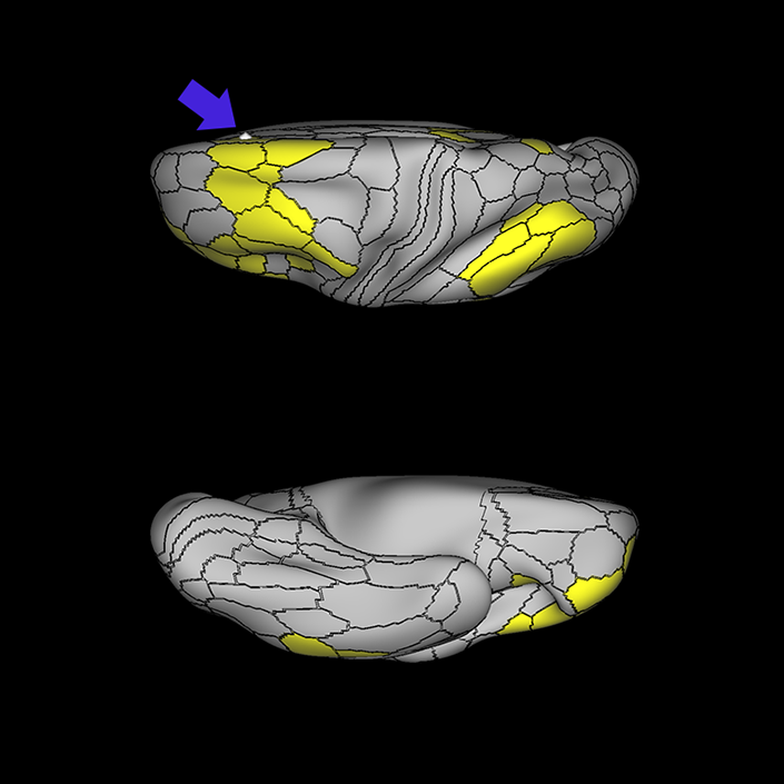
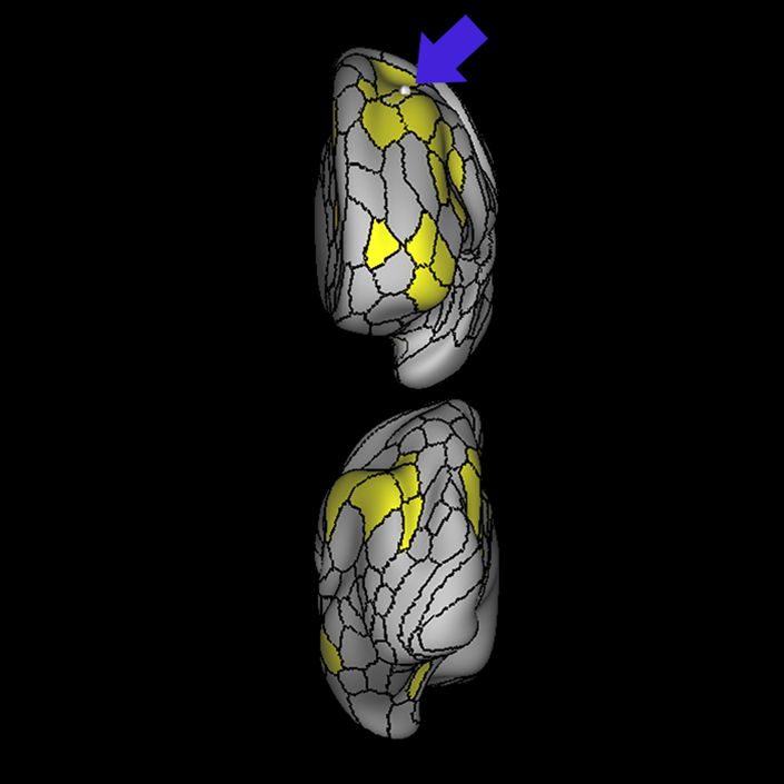
ᐅ SummaryArea SCEF (supplementary and cingulate eye field): part of medial superior frontal gyrus regions. Higher order oculomotor center implicated in appraising all possible oculomotor behaviors to direct primary oculomotor centers in goal- directed behavior. ᐅ Where is it?Area SCEF (supplementary and cingulate eye field) is located in the posterior medial SFG. ᐅ What are its borders?Area SCEF borders area 8BM anteriorly, areas 6ma and SFL superiorly, areas 6mp and 24dd posteriorly, and areas 24dv and p32pr inferiorly. ᐅ What are its functional connections?Area SCEF demonstrates functional connectivity to areas 1, 2, 3a, 3b in the sensory strip, area 4 in the motor strip, areas PEF, FEF, 55b, 6ma, 6mp, 6a, 6r, and 6v in the premotor regions, areas a24prime, p32prime, a32prime, 5mv, and 23c in the middle cingulate regions, areas IFJa, 46, and 9-46d in the lateral frontal lobe areas OP4, OP1, PFcm, 43, FOP1, FOP2, FOP3 FOP4, and FOP5 in the superior insula opercular regions, areas PSL, 52, A4, MI, PoI1 and PoI2 in the lower opercula and Heschl's gyrus regions, area PHT in the temporal lobe, areas AIP, MIP, VIP, LIPd, LIPv, PFop, PF, PFt, IP0, IPS1, 7AL, 7PL, and 7PC, in the lateral parietal lobe, areas 7am and DVT in the medial parietal lobe, area V1, V2, V3, V4 in the medial occipital lobe, areas V3a, V3b, V6, V6a, and V7 in the dorsal visual stream, area FFC of the ventral visual stream, and areas PH, TPOJ2, LO3, MST, and FST of the lateral occipital lobe. ᐅ What are its white matter connections?Area SCEF is structurally connected to the contralateral hemisphere and thalamus. Contralateral connections course through the body of the corpus callosum to end at SCEF, 8BL, SFL and 8BM. Thalamic projections travel through the ventral thalamus to the brainstem (Figure 32). Local short association bundles connect with SF, 8BM, SFL and 8BL. ᐅ What is known about its function?Area SCEF is a higher order oculomotor center implicated in appraising all possible oculomotor behaviors to direct primary oculomotor centers in goal-directed behavior. |
|
A: lateral-medial
B: anterior-posterior
C: superior-inferior
DTI image |