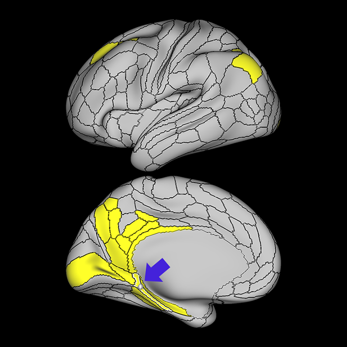
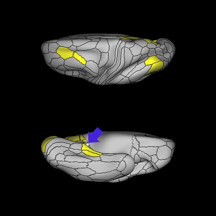
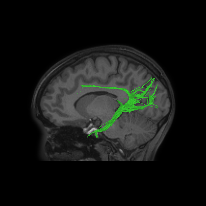
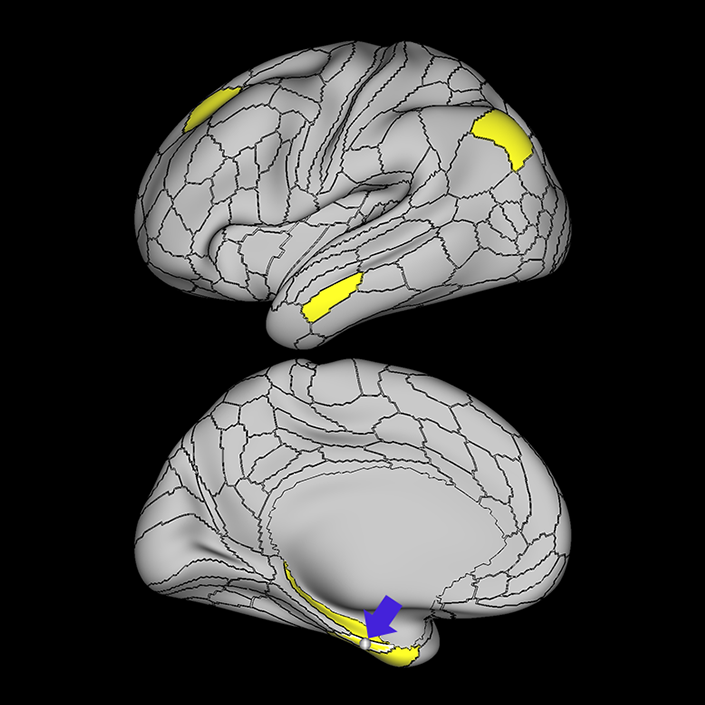
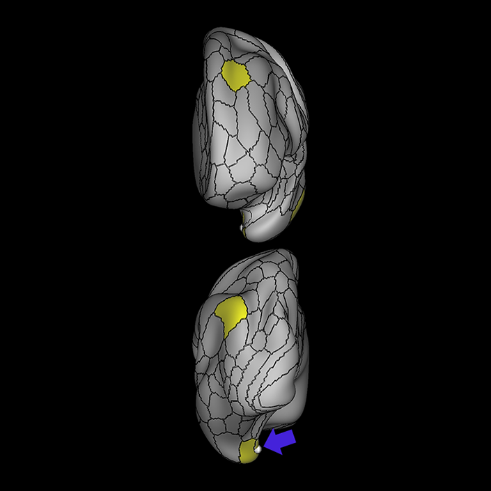
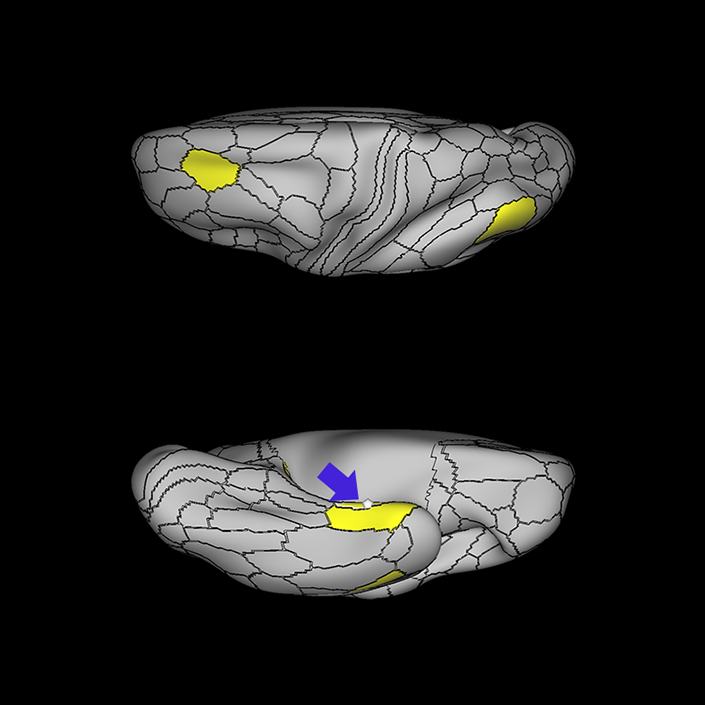
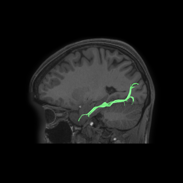
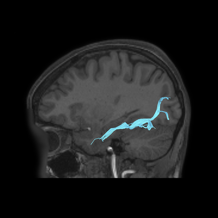
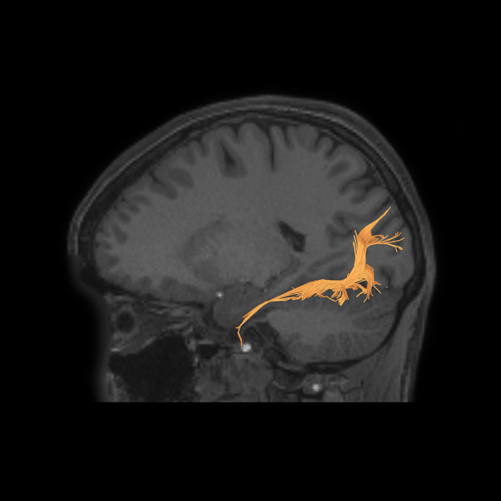
ᐅ SummaryArea EC (entorhinal cortex): part of the temporal lobe regions. Thought to be involved in both the rapid encoding of new associations and in the consolidation of memory in connection with the medial prefrontal cortex. It is less activated in primary task contrasts, ie completed tasks vs baseline fixation, and activated rather than deactivated in working memory of body images. ᐅ Where is it?Area EC (entorhinal cortex) is found on the medial posterior surface of the uncus. ᐅ What are its borders?Area EC borders the presubiculum posteriorly, as well as PHA1. Its inferior and anterior neighbor is PeEC. ᐅ What are its functional connections?Area EC demonstrates functional connectivity to area 8ad in the frontal lobe, areas TE1a, PeEc, and the hippocampus in the temporal lobe, and area PGs in the parietal lobe. ᐅ What are its white matter connections?Area EC is structurally connected to the cingulum. Cingulum fibers span the entire cingulate cortex to terminate anteriorly at a24pr and p24. There are posterior projections from the cingulum that travel toward the parieto-occipital sulcus to terminate at DVT and POS2. ᐅ What is known about its function?The entorhinal cortex (EC) is thought to be involved in both the rapid encoding of new associations and in the consolidation of memory in connection with the medial prefrontal cortex. Relative to the PeEC which borders the region inferiorly and medially, the EC contains more myelin, is thinner, and has different functional connectivity. It is less activated in primary task contrasts, i.e. completed tasks vs baseline fixation, and activated rather than deactivated in working memory of body images. Relative to area PHA1 posteriorly, the EC contains more myelin, is thinner, and is less activated during language tasks and theory of mind tasks |
|
A: lateral-medial
B: anterior-posterior
C: superior-inferior
DTI image |
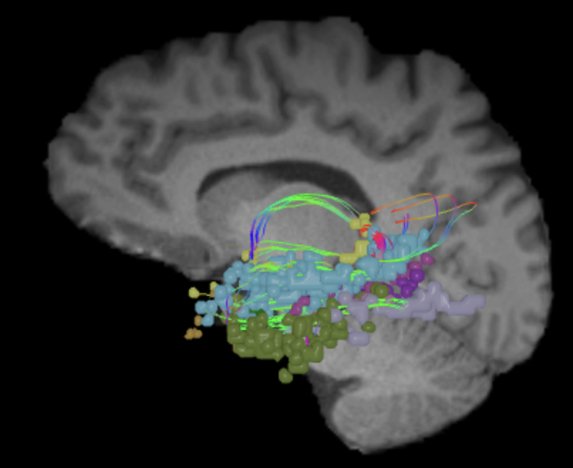
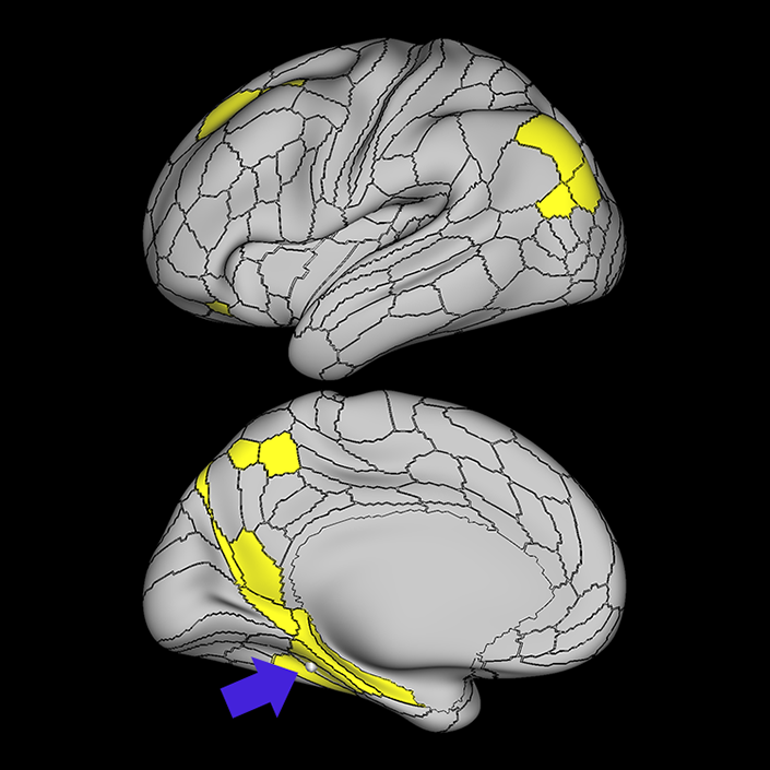
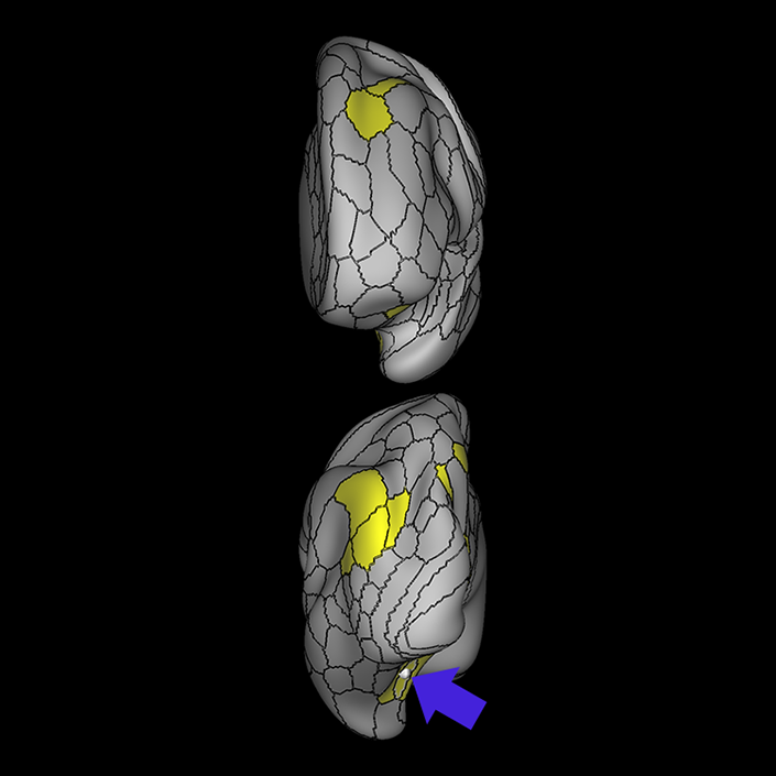
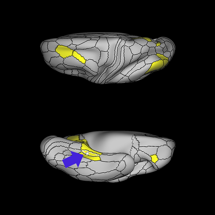
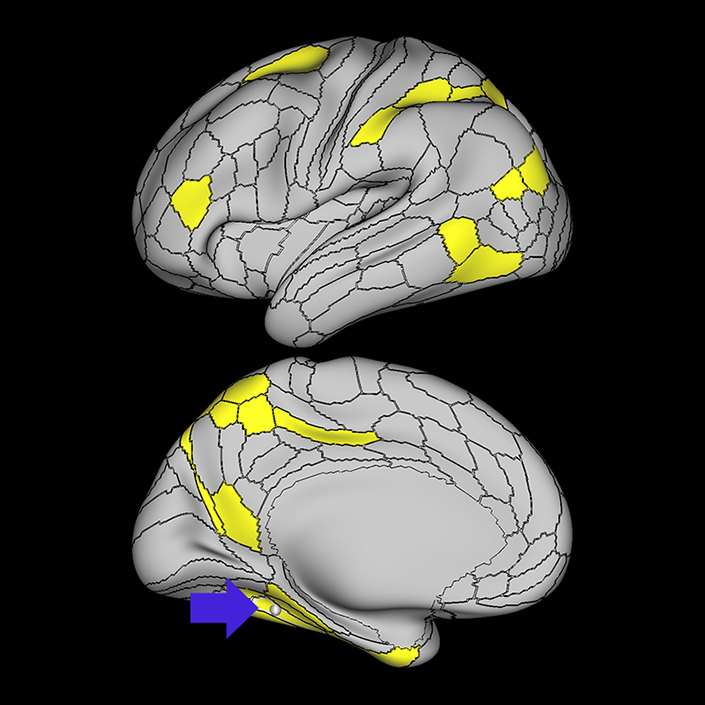
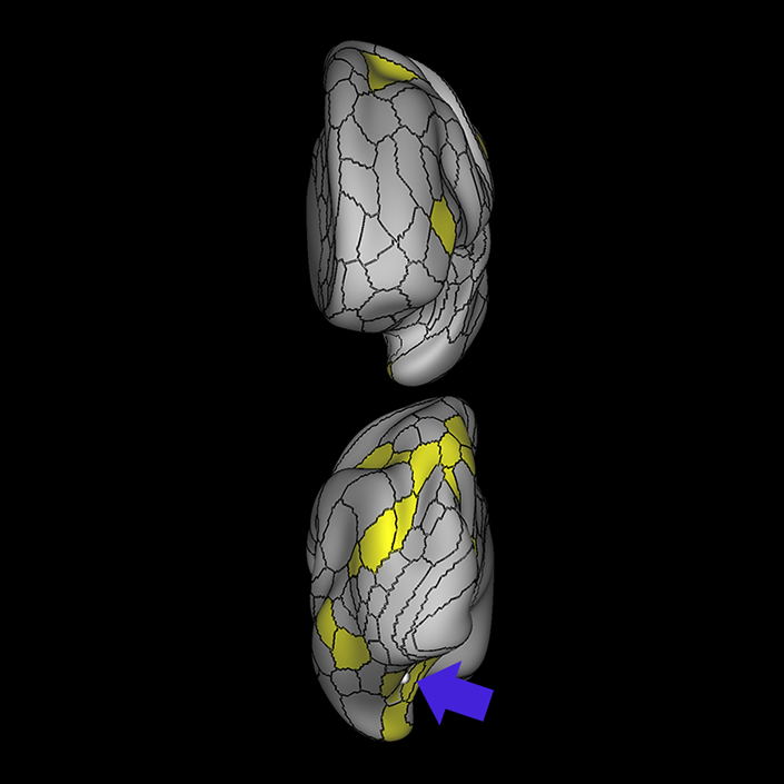
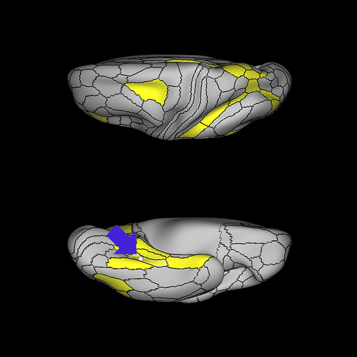
ᐅ SummaryArea PeEc (perirhinal ectorhinal cortex): part of the temporal lobe regions. The perirhinal cortex: contributes to declarative memories transmitted between cortical areas and the hippocampus; adds semantic knowledge to aid in item identification; integrates item information with spatio-temporal information; transmits this data to the hippocampus via the EC. ᐅ Where is it?Area PeEC (perirhinal ectorhinal cortex) is found on the anterior portions and inferior surface of the uncus extending to the collateral sulcus. ᐅ What are its borders?Area PeEC borders areas TGv and TGd anteriorly and TF laterally. Its posterior borders are PHA2 and PHA3. ᐅ What are its functional connections?Area PeEC demonstrates functional connectivity to area IFSa the frontal lobe, areas EC, TF, PHA2, and PHA3 in the temporal lobe, area IP0 in the parietal lobe, and area PH in the occipital lobe. ᐅ What are its white matter connections?Area PeEC is structurally connected to the ILF. ILF projections travel through the inferior temporal lobe to end at PH, TPOJ3 and MT. In some individuals there are fibers that parallel the ILF to terminate at the medial occipital lobe at V1 ᐅ What is known about its function?The perirhinal cortex contributes to declarative memories transmitted between cortical areas and the hippocampus. The perirhinal cortex region adds semantic knowledge to aid in item identification. In addition, the perirhinal cortex integrates item information with spatio-temporal information and transmits this data to the hippocampus via the entorhinal cortex. The HCP authors were unable to reliably separate the perirhinal cortex and ectorhinal cortex, and therefore combined them as a single region. Area PeEC can be distinguished from neighboring regions based on increased activation in working memory primary task contrasts and increased activation in selective recognition of faces. Due to its particular affinity for facial recognition tasks compared to its neighbors, the HCP authors hypothesize that the region may be the site of the anterior temporal face patch |
|
A: lateral-medial
B: anterior-posterior
C: superior-inferior
DTI image |


ᐅ SummaryArea PHA1 (parahippocampal area 1): part of the temporal lobe regons. Involved in visuospatial processing and episodic memory by processing contextual information. The anterior parahippocampal cortex is involved in encoding information without regard for stimulus category (scenes vs objects) or modality (word vs picture) and interfaces with the hippocampus, retrosplenial, and perirhinal memory systems, while the posterior parahippocampal cortex is involved with pictorial scene analysis, namely processing spatial features of visual scenes. ᐅ Where is it?Area PHA1 (parahippocampal area 1) is a long thin area on the medial portion of the parahippocampal gyrus. ᐅ What are its borders?Area PHA1 borders area PreS superiorly and PHA2 inferiorly. Its posterior border is VMV1 and its anterior border is EC. ᐅ What are its functional connections?Area PHA1 demonstrates functional connectivity to areas 10r and 8ad in the frontal lobe, areas PHA2, PHA3, VMV1 PreS, the hippocampus in the temporal lobe, and areas ProS, POS1, PGp, and PGs in the parietal lobe. ᐅ What are its white matter connections?Area PHA1 is connected to local parcellations. Local anterior fibers connect to PeEc. Local posterior fibers connect to PH. The exact terminations of the local connections are inconsistent across individuals. ᐅ What is known about its function?The parahippocampal cortex is involved in visuospatial processing and episodic memory by processing contextual information. The anterior parahippocampal cortex is involved in encoding information without regard for stimulus category (scenes vs objects) or modality (word vs picture) and interfaces with the hippocampus,retrosplenial, and perirhinal memory systems while the posterior parahippocampal cortex is involved with pictorial scene analysis, namely processing spatial features of visual scenes. Like both PHA2 and PHA3, PHA1 is activated in the PLACE-AVG contrast and deactivated in the FACE-AVG contrast, suggesting a role in place/scene recognition rather than face recognition. However, PHA1 is less deactivated in face recognition than PHA2 and PHA3 inferiorly. |
|
A: lateral-medial
B: anterior-posterior
C: superior-inferior
DTI image |
ᐅ SummaryArea PHA2 (parahippocampal area 2): part of the temporal lobe regions. Involved in visuospatial processing and episodic memory by processing contextual information. The anterior parahip- pocampal cortex is involved in encoding information without regard for stimulus category (scenes vs objects) or modality (word vs picture) and interfaces with the hippocampus, retrosplenial, and perirhinal memory systems, while the posterior parahippocampal cortex is involved with pictorial scene analysis, namely processing spatial features of visual scenes. ᐅ Where is it?Area PHA2 (parahippocampal area 2) is found on parahippocampal gyrus, primarily on its inferior surface, just adjacent to the collateral sulcus. ᐅ What are its borders?Area PHA2 is a long thin anterior posterior area between PHA1 superiorly and PHA3 inferiorly. It has a small porterior border with VMV2. ᐅ What are its functional connections?Area PHA2 demonstrates functional connectivity to areas 8AD, 47m, and i6-8 in the frontal lobe, areas PHA1, PHA3, PreS, and the hippocampus in the temporal lobe, areas ProS, 7pm, PCV, DVT, POS1, IP0, PGp, and PGs in the parietal lobe, and area TPOJ3 in the occipital lobe. ᐅ What are its white matter connections?Area PHA2 is structurally connected to the ILF. There are anterior projections from the ILF that terminate at PeEc. The posterior terminations of the ILF terminate at V1, V2 and V3. The exact terminations of the local connections are inconsistent across individuals. ᐅ What is known about its function?The parahippocampal cortex is involved in visuospatial processing and episodic memory by processing contextual information. The anterior parahippocampal cortex is involved in encoding information without regard for stimulus category (scenes vs objects) or modality (word vs picture) and interfaces with the hippocampus, retrosplenial, and perirhinal memory systems while the posterior parahippocampal cortex is involved with pictorial scene analysis, namely processing spatial features of visual scenes. Like both PHA1 and PHA3, PHA2 is activated in the PLACE-AVG contrast and deactivated in the FACE-AVG contrast, suggesting a role in place/scene recognition rather than face recognition. Area PHA2 is more deactivated than PHA1 in facial recognition tasks. |
|
A: lateral-medial
B: anterior-posterior
C: superior-inferior
DTI image |
ᐅ SummaryArea PHA3 (parahippocampal area 3): part of the temporal lobe regions. Involved in visuospatial processing and episodic memory by processing contextual information. The anterior parahippocampal cortex is involved in encoding information without regard for stimulus category (scenes vs objects) or modality (word vs picture) and interfaces with the hippocampus, retrosplenial, and perirhinal memory systems, while the posterior parahippocampal cortex is involved with pictorial scene analysis, namely processing spatial features of visual scenes. ᐅ Where is it?Area PHA3 (parahippocampal area 3) is located in the parahippocampal gyrus, primarily inside the collateral sulcus ᐅ What are its borders?Area PHA3 borders PHA2 superiorly, and VVC (ventral visual complex) and TF inferiorly. Its posterior border is with VMV2 and VMV3, and its anterior border is PeEC. ᐅ What are its functional connections?Area PHA3 demonstrates functional connectivity to areas 6a and IFSa in the frontal lobe, areas PHT, PHA1, PHA2, VMV2 and PeEc in the temporal lobe, areas 23c, 7pl, 7am, 7pm, PCV, DVT POS1, MIP, LIPd, AIP, PGp, and PFt in the parietal lobe, and areas TPOJ3 and VVC in the occipital lobe. ᐅ What are its white matter connections?Area PHA3 is structurally connected to the ILF. There are anterior projections from the ILF that terminate at TGd. The posterior terminations of the ILF terminate at VVC and FFC. There are fibers that run parallel to the ILF and terminate at the occipital lobe at V1, V2 and V6. The exact terminations of the local connections are inconsistent across individuals. ᐅ What is known about its function?The parahippocampal cortex is involved in visuospatial processing and episodic memory by processing contextual information. The anterior parahippocampal cortex is involved in encoding information without regard for stimulus category (scenes vs objects) or modality (word vs picture) and interfaces with the hippocampus, retrosplenial, and perirhinal memory systems while the posterior parahippocampal cortex is involved with pictorial scene analysis, namely processing spatial features of visual scenes. Relative to PHA2, area PHA3 demonstrates greater activity in tool-related recognition tasks. |
|
A: lateral-medial
B: anterior-posterior
C: superior-inferior
DTI image |
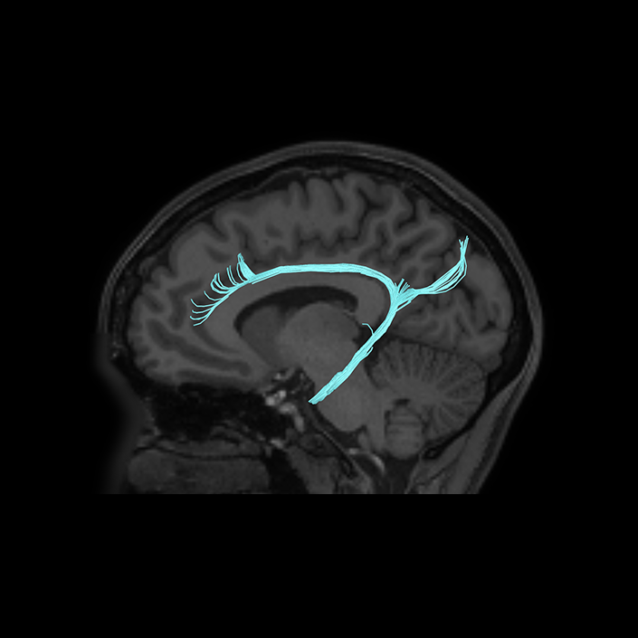
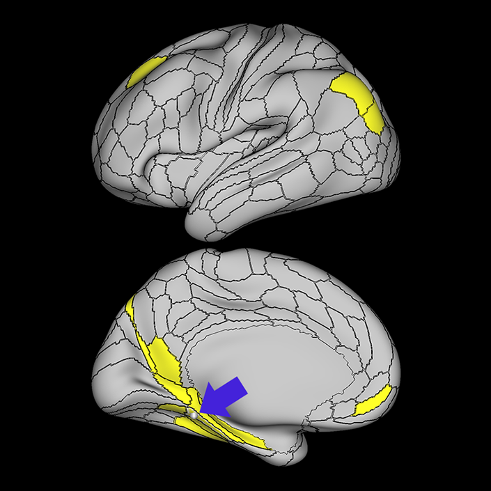
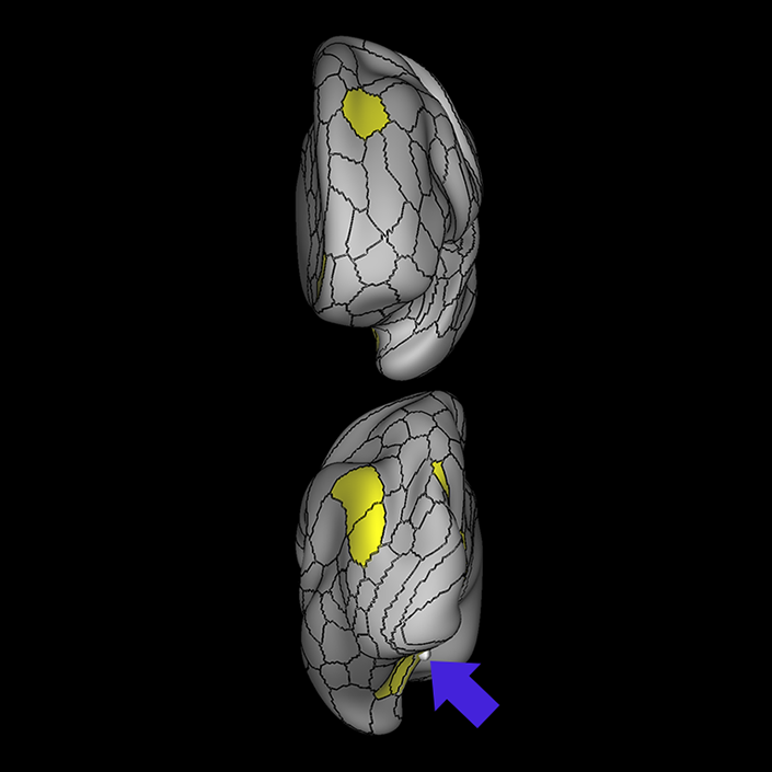
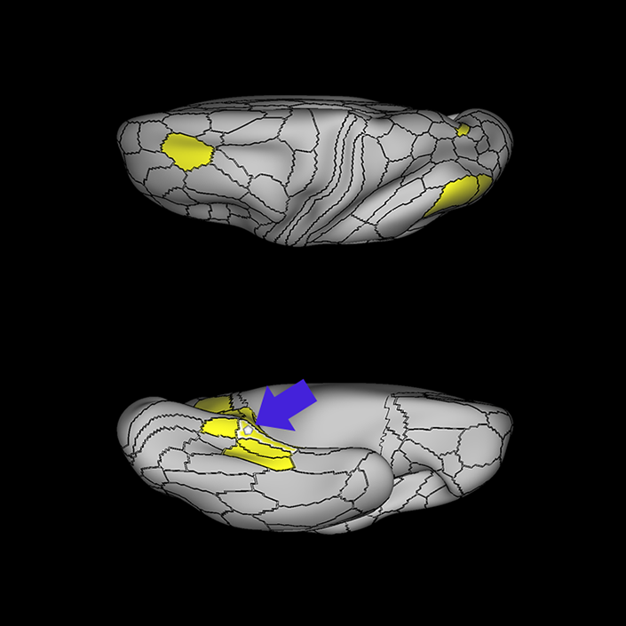
ᐅ SummaryArea PreS: (presubiculum) part of the medial temporal areas. Region of the hippocampus that primate studies suggest is involved in the processing of spatial information. Demonstrates less activity during tasks related to working memory, language processing, and theory of mind. Relative to PHA1, PreS shows greater activity during motor tasks. ᐅ Where is it?Area PreS is found on the posterior superior surface of the parahippocampal gyrus. ᐅ What are its borders?Area PreS borders the hippocampus medially, and the entrorhinal cortex anteriorly. Its posterior border is made up of RSC (retrosplenial cortex) and the ProS (prostriate region) (which are discussed in other sections. PHA1 is its inferior border ᐅ What are its functional connections?Area PreS demonstrates functional connectivity areas 8AD and i6-8 in the frontal lobe, areas PHA1, PHA2, and the hippocampus in the temporal lobe, areas RSC, Pros, d23ab, v23ab, 31a, 31pv, 7m, 7pm, POS1, POS2, IP1, and PGs in the parietal lobe, and area V1 in the occipital lobe ᐅ What are its white matter connections?Area PreS is structurally connected to the cingulum, precuneus and occipital lobe. Cingulum projections run superior to the corpus callosum to end at anterior cingulate cortex and frontal lobe parcellations a24, 9m, 10d and p32. There arePreS fibers that project posteriorly to end at occipital and precuneus areas V1, V2, V6, POS1, POS2 and 7m. Local short association fibers are connected to EC and PeEc. ᐅ What is known about its function?The presubiculum lies medial to the subiculum - a region of the hippocampus which primate studies suggest is involved in the processing of spatial information. Area PreS contains more myelin than the hippocampal cortex, and relative to PHA1 inferiorly, contains more myelin, is thinner, and demonstrates less activity during tasks related to working memory, language processing, and theory of mind. Relative to PHA1, PreS shows greater activity during motor tasks. |
|
A: lateral-medial
B: anterior-posterior
C: superior-inferior
DTI image |