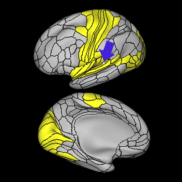
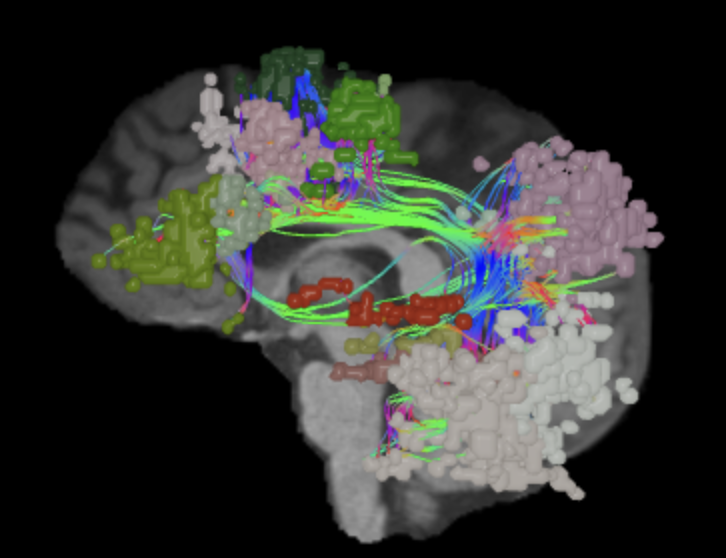
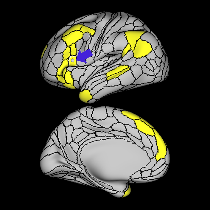
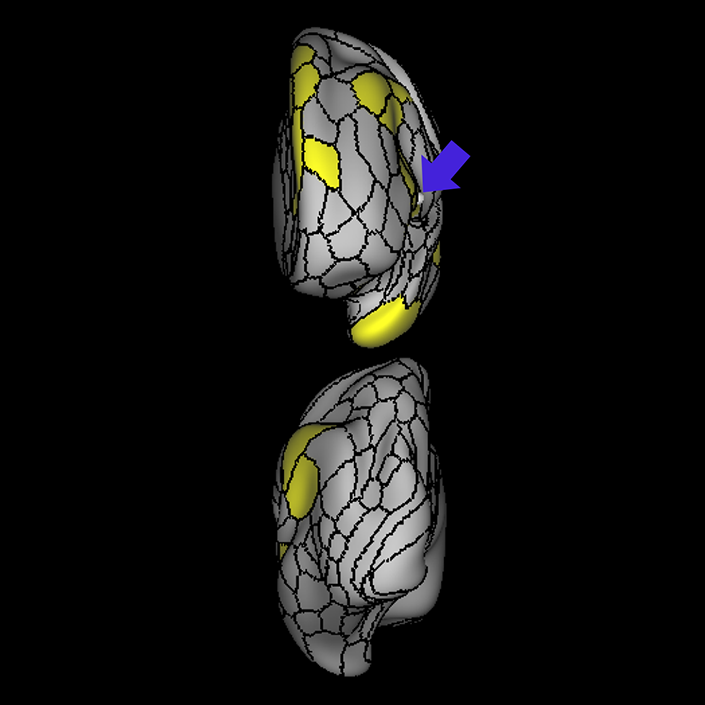
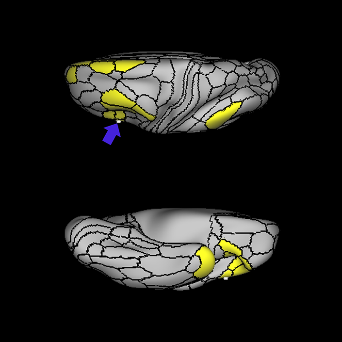
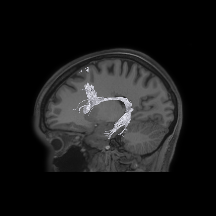
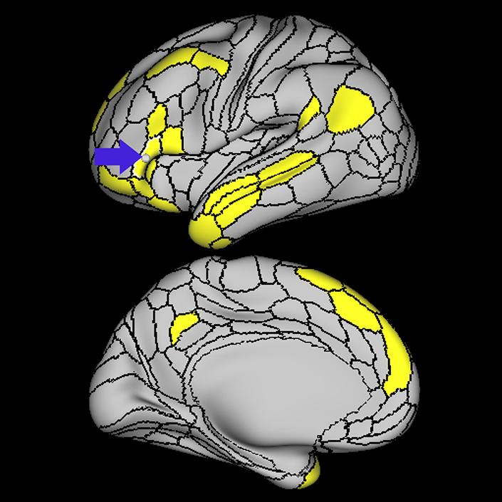
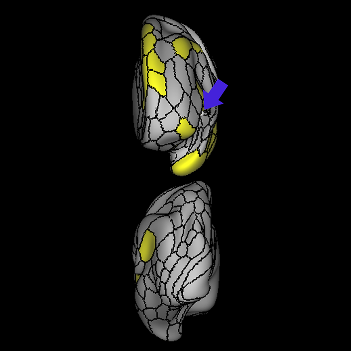
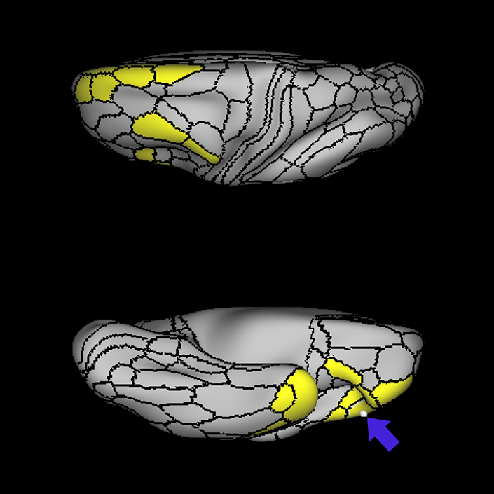
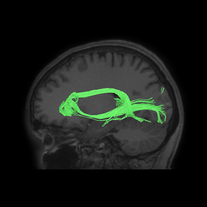
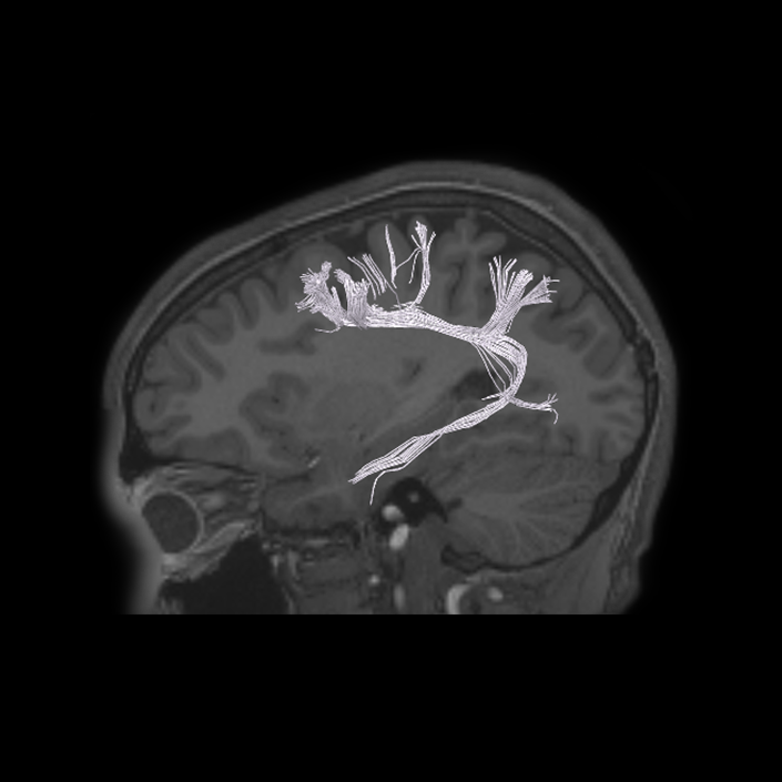
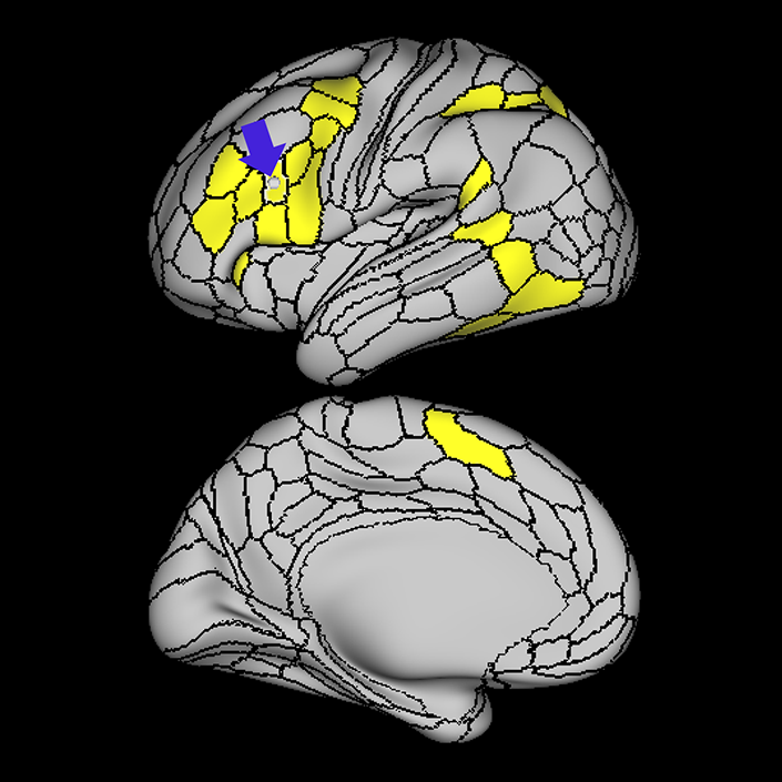
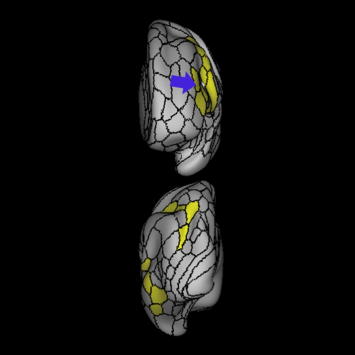
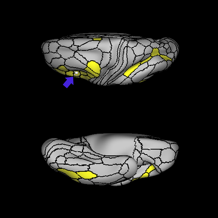
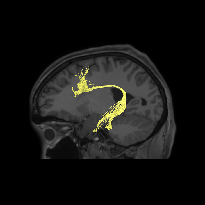
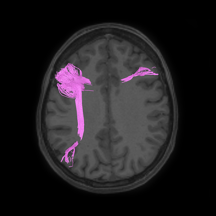
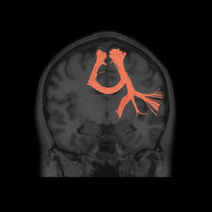
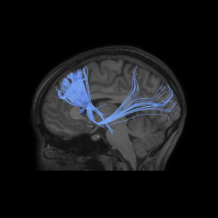
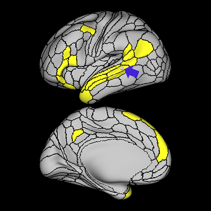
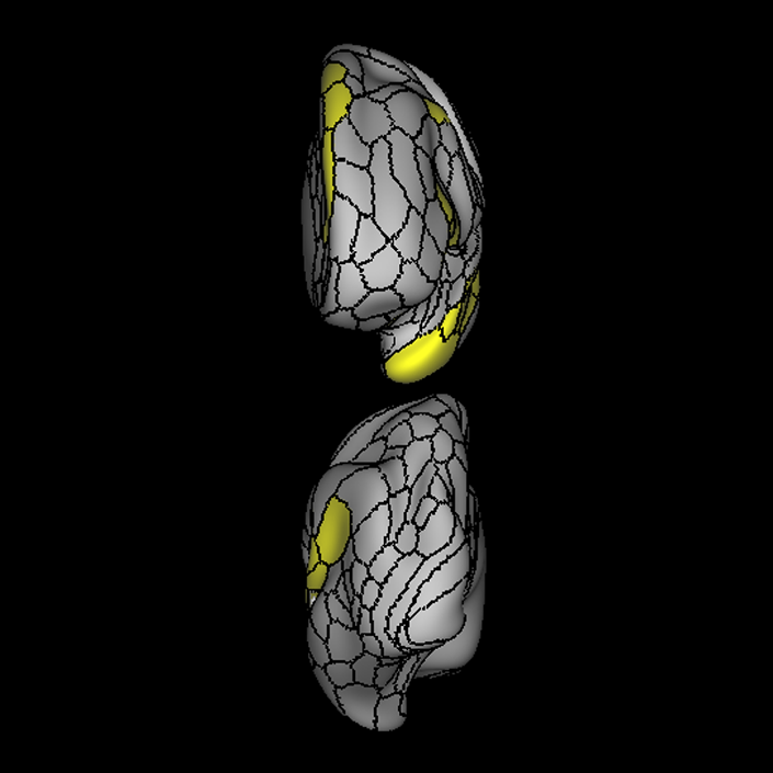
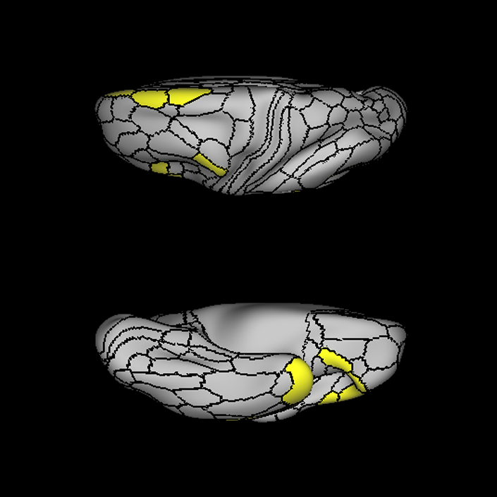
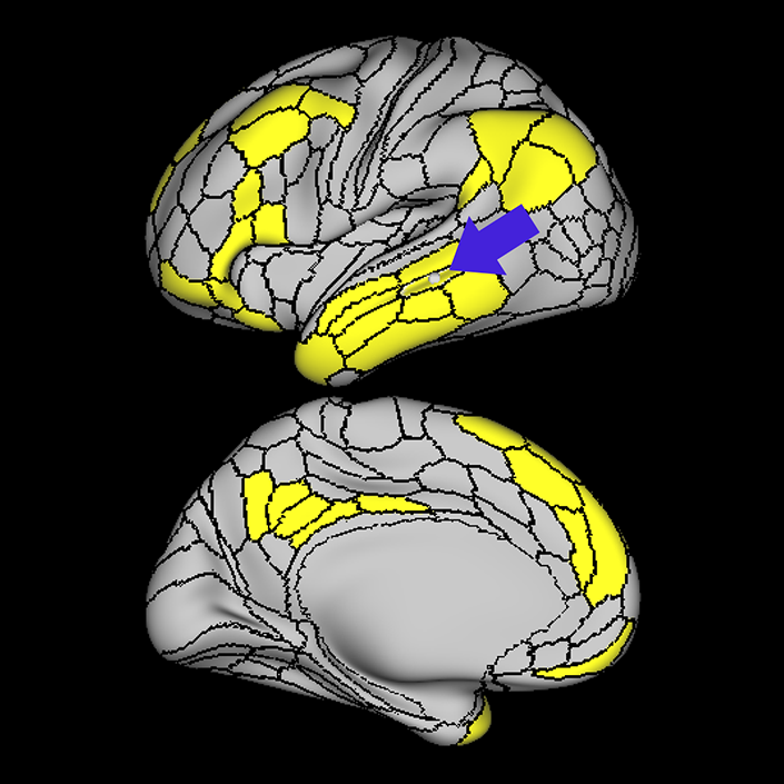
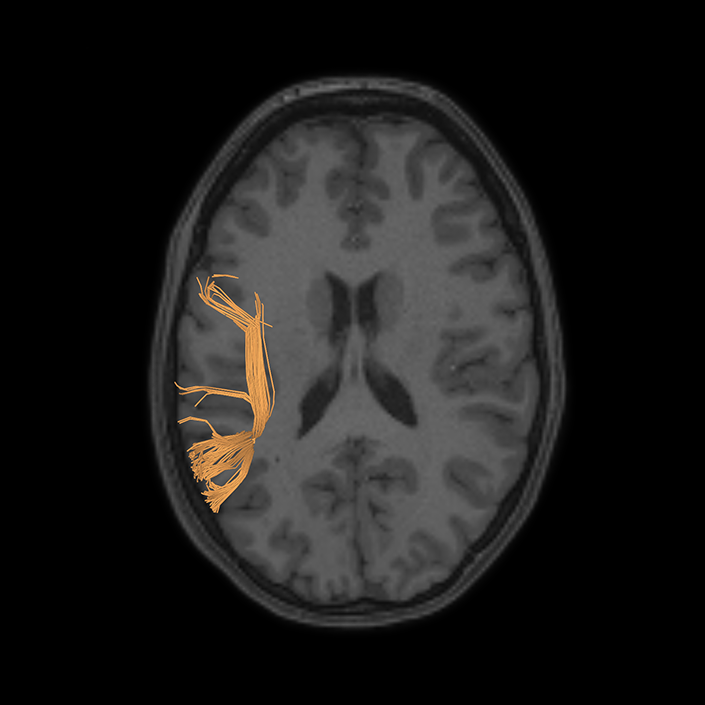
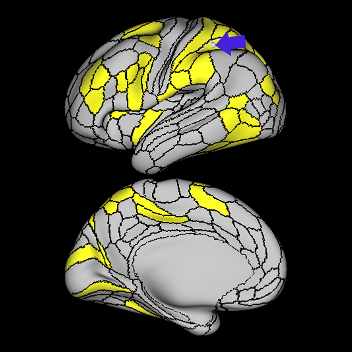
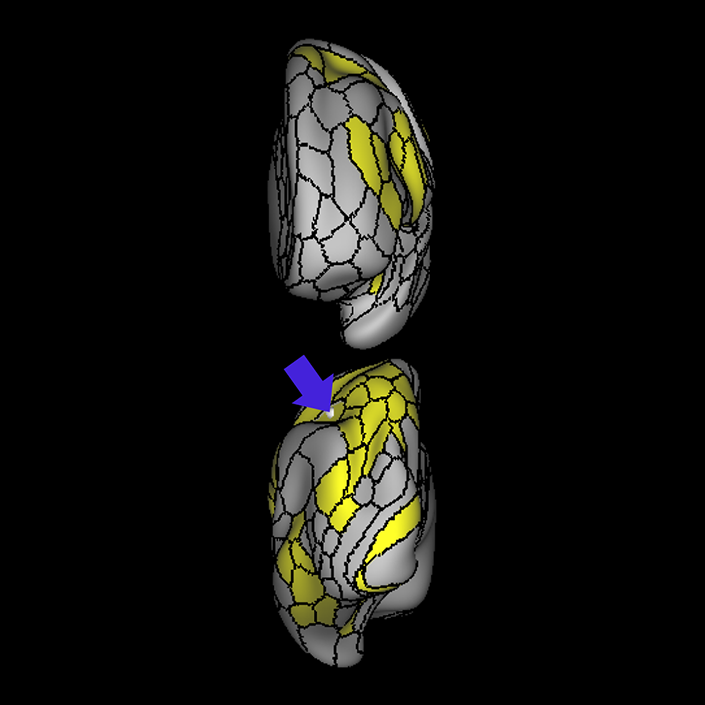
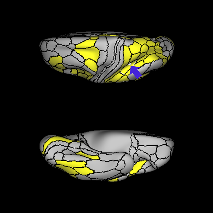
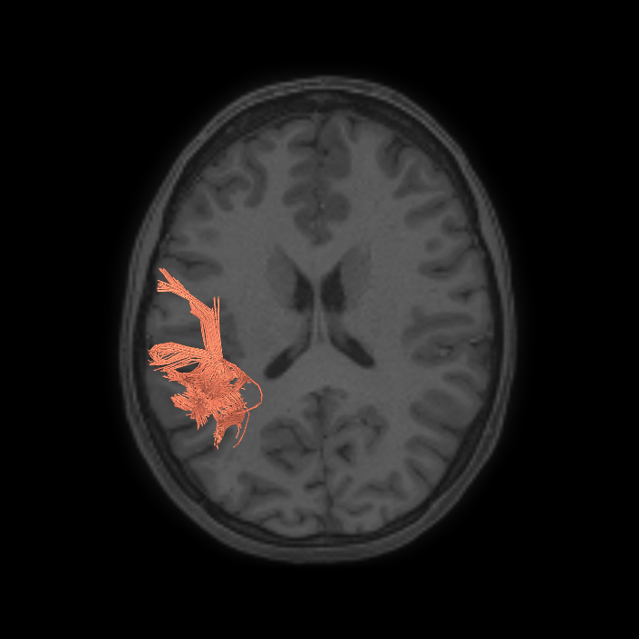
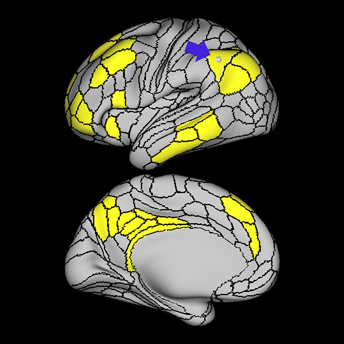
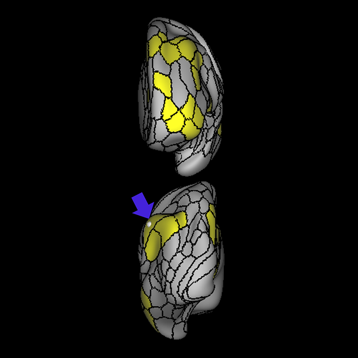
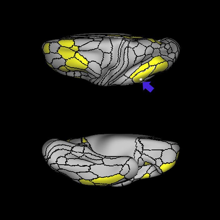
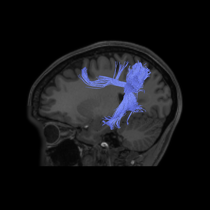
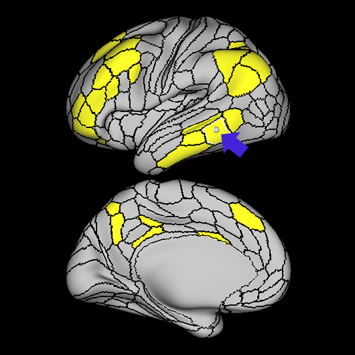
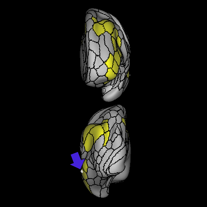
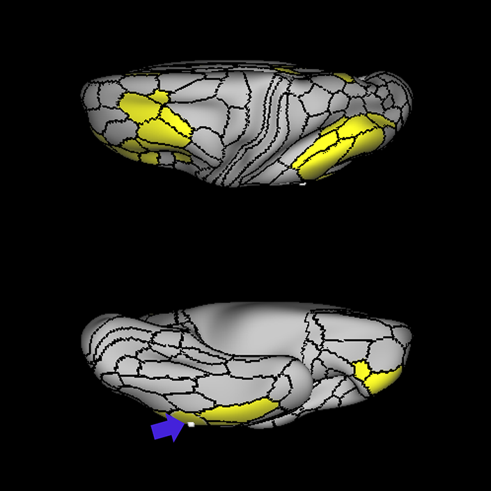
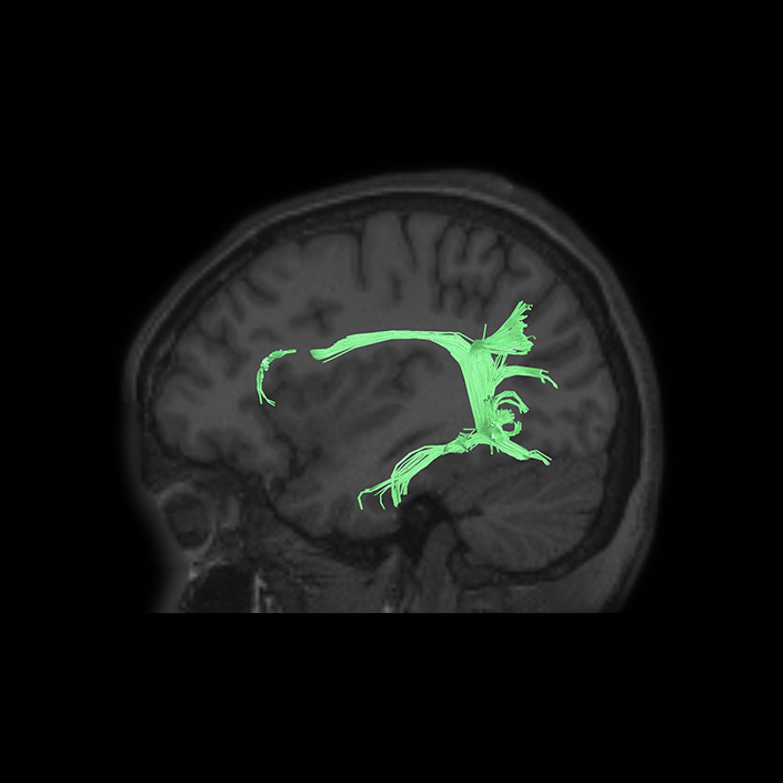
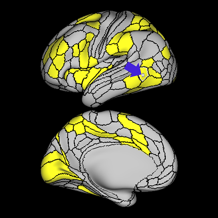
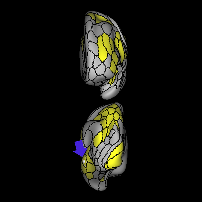
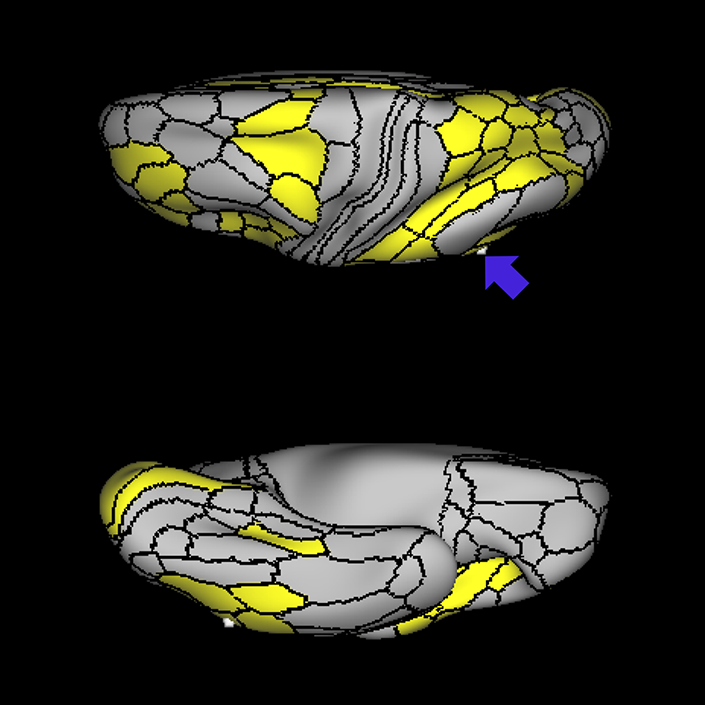
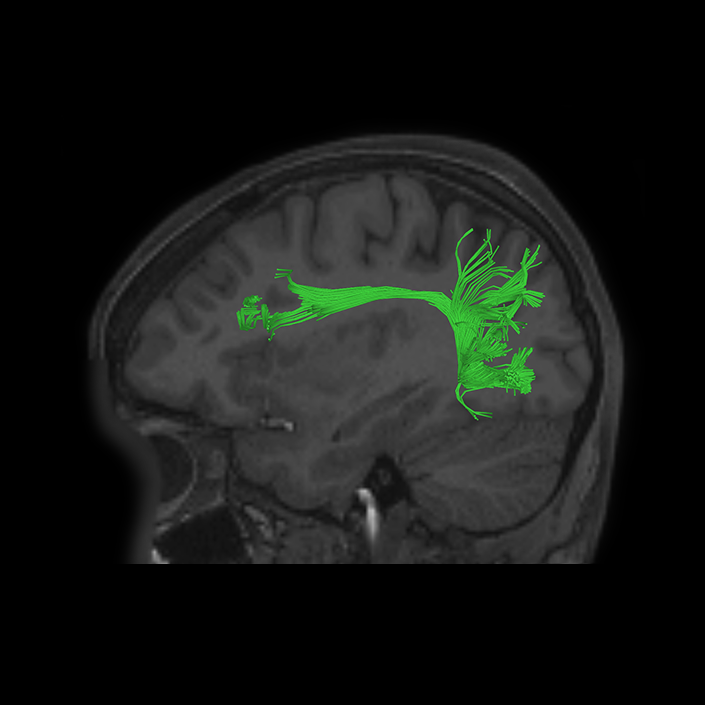
ᐅ SummaryArea 44: part of the inferior frontal gyrus of the lateral frontal lobe. Translates abstract and intentional information in the prefrontal cortex to more detailed representations to help guide the production of verbal and manual actions. In addition to its known association with Broca's area, is sometimes represented as part of Broca's complex ᐅ Where is it?Area 44 is at the posterior most part of the inferior frontal gyrus. It is the anterior bank of pars opercularis of the IFG. ᐅ What are its borders?Area 44 borders area 45 anteriorly and area 6r posteriorly. Area 8C is its medial border and its inferior border is wedged between then upper borders of Areas 6R and 6V. Its superior edge borders IFSp and IFJa. Its opercular surface is FOP4. ᐅ What are its functional connections?Area 44 demonstrates functional connectivity to areas SFL, IFSp, IFJa, 45, 47s, 47L, 9a, 9m, 8AV, 8BL and 8C in the dorsolateral frontal lobe, area 8BM in the medial frontal lobe, area 55b in the premotor areas, areas FOP5, AVI and PSL in the insula- opercular region, areas TGd, STSdp and STSvp in the temporal lobe, areas PFm, and PGi in the inferior parietal lobe, and no areas in the medial parietal lobe. ᐅ What are its white matter connections?Area 44 is structurally connected to the arcuate/SLF and the FAT. Connections with the arcuate/SLF project posteriorly and wrap around the Sylvian fissure to the middle temporal gyrus to end at TE1a and TE1m. There are also projections from the arcuate/SLF before it terminates to parcellations A5 and STSdp. The majority of the inferior connections of the frontal aslant tract end at 44, the tract is connected superiorly to superior frontal gyrus parcellations SFL, 6ma and s6-8. Local short association bundles are connected with 45 and 8C. White matter tracts from 44 in the right hemisphere have less consistent connections with the arcuate/SLF. ᐅ What is known about its function?Area 44 translates abstract and intentional information in the prefrontal cortex to more detailed representations to help guide the production of verbal and manual actions. Area 44, in addition to its known association with Broca's area, is sometimes represented as part of "Broca's complex", including Brodmann Areas 45, 46, 47 and the mesial supplementary motor area of 6, which contribute to a frontal-subcortical circuit. The right pars opercularis has also been implicated in cognitive inhibition in the overall context of working memory. |
|
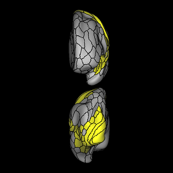
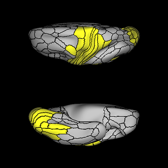
A: lateral-medial
B: anterior-posterior
C: superior-inferior
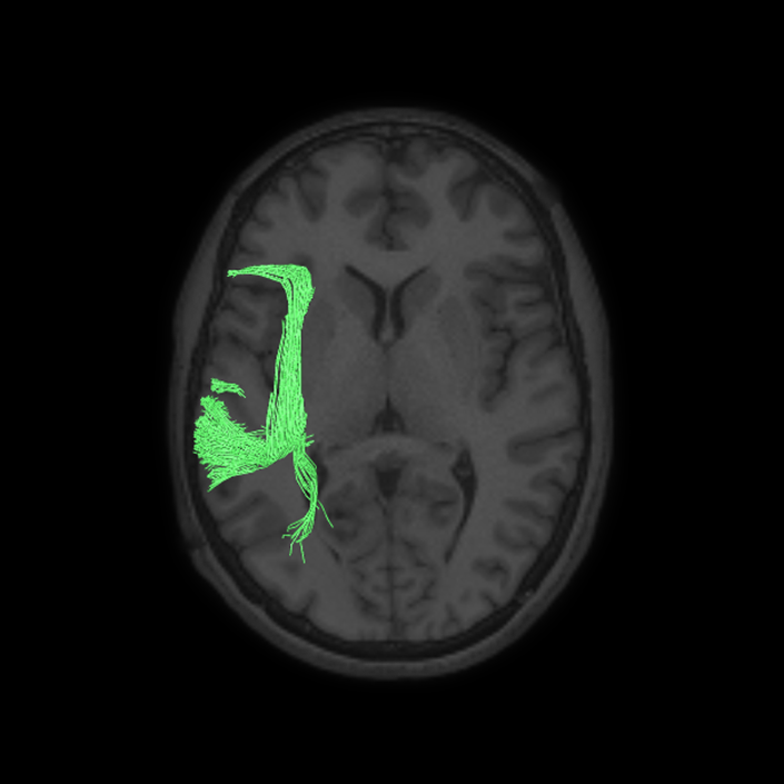
DTI image |
ᐅ SummaryArea 45: part of the inferior frontal gyrus of the lateral frontal lobe. In addition to its known association with Broca's area, is sometimes represented as part of Broca's complex ᐅ Where is it?Area 45 is the lateral surface of pars triangularis of the IFG. ᐅ What are its borders?Area 45 borders area 47L anteriorly and area 44 posteriorly. Its superior edge borders area p47r, IFSa, and IFSp. Its opercular surface is conveniently named FOP5 ᐅ What are its functional connections?Area 45 demonstrates functional connectivity to areas SFL, IFSp, 44, a47r, 47s, 47L, 9a, 9p, 9m, 8AV, and 8BL in the dorsolateral frontal lobe, area 8BM in the medial frontal lobe, area 55b in the premotor areas, areas FOP5, and PSL in the insula-opercular region, areas TGd, TGv, TE1a, STSva, STSdp and STSvp in the temporal lobe, area PGi in the inferior parietal lobe, and area 31pd in the medial parietal lobe. ᐅ What are its white matter connections?Area 45 is structurally connected to the arcuate/SLF and IFOF. However, arcuate/SLF connections are not consistent across individuals. Connections with the arcuate/SLF project posteriorly and wrap around the Sylvian fissure to the middle temporal gyrus to end at TE1p. There are also projections from the arcuate/SLF before it terminates to parcellations A4 and PBelt. IFOF connections travel from 45 through the extreme/external capsule and continue posteriorly through the temporal lobe to end at occipital lobe parcellations V1, V2, V3 and V4. Local short association bundles connect with 44 and FOP4. ᐅ What is known about its function?Area 45, in addition to its known association with Broca's area, is sometimes represented as part of "Broca's complex", including Brodmann Areas 45, 46, 47 and the mesial supplementary motor area of 6, which contribute to a frontal-subcortical circuit. |
|
A: lateral-medial
B: anterior-posterior
C: superior-inferior
DTI image |
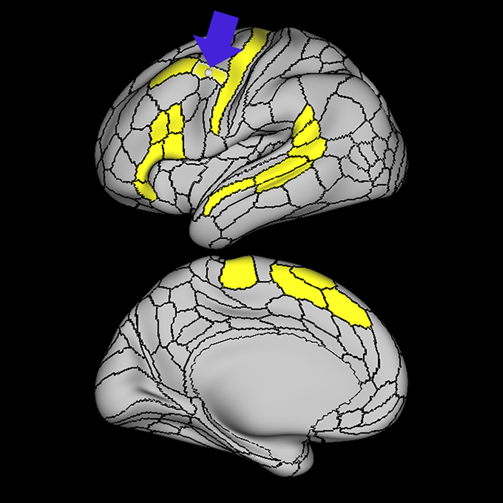
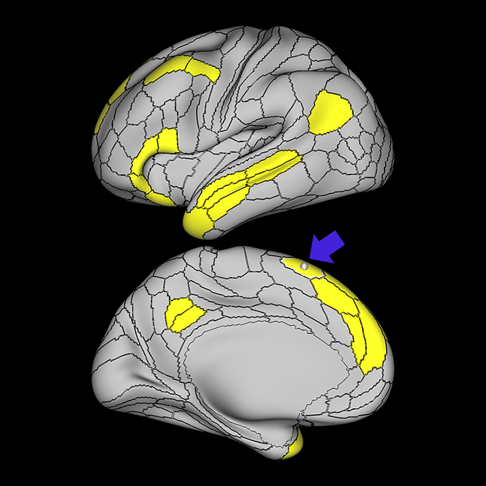
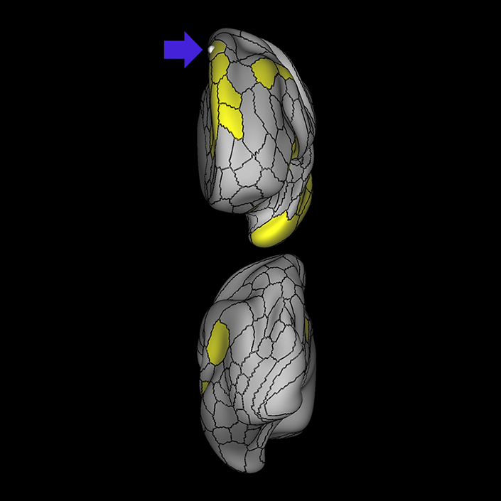
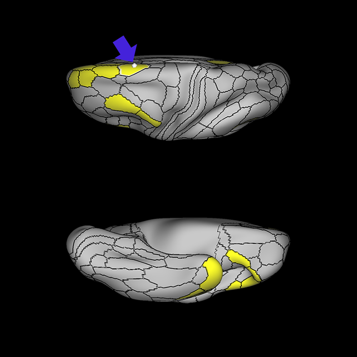
ᐅ SummaryArea 55b: part of the premotor areas. Relatively uncharacterized regions. In 1956, one of the only studies to characterize this regions concluded that the area played a role in language processing. ᐅ Where is it?Area 55b is located on the anterior half of the precental gyrus, approximately half way down its length along the convexity, just inferior to FEF. It also forms the adjacent floor of the precentral sulci and straddles slightly onto the posterior edge of the middle frontal gyrus. ᐅ What are its borders?Area 55b borders area FEF superiorly and PEF and area 6v inferiorly. Area 4 is its posterior border and areas 8AV and 8C form its anterior border across the precentral sulcus. ᐅ What are its functional connections?Area 55b demonstrates functional connectivity to area 4 in the motor strip, areas SCEF and SFL in the premotor areas, areas IFSp, IFJa, 8AV, 44, 45, and 47L in the lateral frontal lobe, areas STSda and STSdp in the temporal lobe, areas PSL and STV in the posterior opercular cortices, and area TPOJ1 in the lateral occipital lobe. ᐅ What are its white matter connections?Area 55b is structurally connected to the contralateral hemisphere and the superior longitudinal fasciculus. Contralateral connections course through the body of the corpus callosum to 6ma, 6a and 6mp. Connections with the superior longitudinal fasciculus connect 55b to parcellations PHT and PFm, and this tract terminates eventually in the temporal lobe at TGd. Local short association fibers connect with 8Av, 8C, IFJp, 3a, 3b and PEF. ᐅ What is known about its function?Area 55b is a relatively uncharacterized region. In 1956, one of the only studies to characterize this region concluded that the area played a role in language processing. |
|
A: lateral-medial
B: anterior-posterior
C: superior-inferior
DTI image |
ᐅ SummaryArea IFJa (inferior frontal junction, anterior): part of the lateral frontal lobe. Areas in the midventrolateral prefrontal cortex interact with posterior areas of the brain to retrieve specific auditory memories. The inferior frontal junction, in particular, serves as an important crossroads between bottom-up and top-down processing in the lateral prefrontal cortex. ᐅ Where is it?Area IFJA is located in the posterior portion of the inferior frontal sulcus. It comprises part of the inferior bank of the MFG in its upper portions. It is roughly superior to the pars opercularis portion of the inferior frontal gyrus. ᐅ What are its borders?Area IFJa borders area IFSp anteriorly and IFJp posteriorly. Its inferior border is area 44 and its superior border is area 8C. ᐅ What are its functional connections?Area IFJa demonstrates functional connectivity to areas 44, IFSa, IFSp, IFJp, and p9-46v in the dorsolateral frontal lobe, area SCEF in the medial frontal lobe, areas FEF, 55b PEF and 6r in the premotor areas, area FOP5 and PSL in the insular opercular regions, areas PH, PHT, and TE2p in the temporal lobe, areas MIP, TPOJ1, and LIPd in the inferior parietal lobe, and no areas in the medial parietal lobe. ᐅ What are its white matter connections?Area IFJa is structurally connected with the arcuate/SLF and surrounding parcellations. Connections with the arcuate/SLF project posteriorly and wrap around the Sylvian fissure to the middle and inferior temporal gyrus to end at TE1a, TE1m, and TE2a. There are also fibers that project superiorly form IFJa to end at SFL. These fibers are likely portions of the frontal aslant tract which has the majority of its inferior terminations at 44, a neighbor of IFJa. Local short association bundles connect to 8c, IFJa, IFSp, 44 and 8A. ᐅ What is known about its function?Areas in the midventrolateral PFC interact with posterior areas of the brain to retrieve specific auditory memories. The IFJ, in particular, serves as an important crossroads between bottom-up and top-down processing in the lateral prefrontal cortex. |
|
A: lateral-medial
B: anterior-posterior
C: superior-inferior
DTI image |
ᐅ SummaryArea 8C: part of the lateral frontal lobe regions. Within the context of spatial working memory, area 8C is involved in the interpretation of complex visual information and attention. Areas 8 and rostral 6, as part of the posterior dorsolateral frontal areas, are also involved in the maintenance of spatial information. ᐅ Where is it?Area 8C is located at the posterior part of the middle frontal gyrus. It is an anterior- to-posterior band which is lateral to area 8AV. ᐅ What are its borders?Area 8C has area 8AV as its main medial border. Its lateral border is with 3 inferior frontal sulcus areas: IFSp, IFJa, and IFJp. Area 55b and PEF (precentral eyefield) are its posterior borders (and discussed in a different section). Area p9-46v and to a lesser extent area 46 are its anterior neighbors. ᐅ What are its functional connections?Area 8C demonstrates functional connectivity to areas s6-8, i6-8, a9-46v, p9-46v, a10p, 8BL, 8AD, and 8AV in the dorsolateral frontal lobe, areas 8BM and d32 in the medial frontal lobe, areas IFSp, IFJp, a47r, p47r, and 44 in the inferior frontal lobe, area AVI in the insula, areas TE1m, TE1p, TE2a, STSva, and STSvp in the temporal lobe, areas IP1, IP2, LIPd, PFm, PGi and PGs in the inferior parietal lobe, and areas 7pm, 31pv, 31a, POS2, 23d, and d23ab in the medial parietal lobe. ᐅ What are its white matter connections?Area 8C is structurally connected to the arcuate/SLF and the contralateral hemisphere. Contralateral connections travel through the corpus callosum to end at 8C. Connections with the arcuate/SLF project posteriorly and wrap around the sylvian fissure to the posterior temporal lobe to end at PH and PHT. ᐅ What is known about its function?Within the context of spatial working memory, area 8C is involved in the interpretation of complex visual information and attention. Areas 8 and rostral 6, as part of the posterior dorsolateral frontal areas, are also involved in the maintenance of spatial information. |
|
A: lateral-medial
B: anterior-posterior
C: superior-inferior
DTI image |
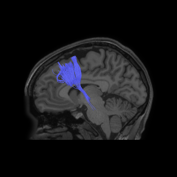
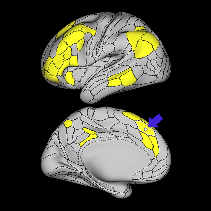
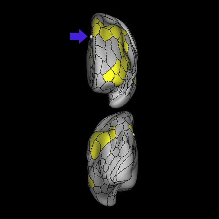
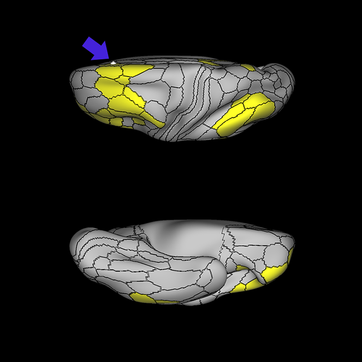
ᐅ SummaryArea SFL: part of the supplementary motor regions. Known to be hemispherically asymmetric. Specifically, the left hemisphere shows more activity when listening to stories and when a participant is matching objects based on a verbal cue. Compared to area 8BL, area SFL shows more activation when listening to a story, matching objects based on verbal cues and in social interaction settings. Compared to area s6-8, area SFL shows more activation in the left hemisphere when individuals listen to a story. In the right hemisphere, area SFL is activated insocial interaction settings and is deactivated during object feature comparison tasks. ᐅ Where is it?Area SFL (superior frontal language area) is located on the posterior medial SFG straddling over the interhemispheric cleft. ᐅ What are its borders?Area SFL borders SCEF inferiorly. Its anterior inferior neighbor is area 8BM and its anterior superior neighbor is area 8BL. Areas 6ma and s6-8 are its lateral neighbors. ᐅ What are its functional connections?Area SFL demonstrates functional connectivity to areas 8BL, 8AV, 9a, 9p, and 9m in dorsolateral frontal lobe, areas 8BM, d32, areas 44, 45, 47L, and 47s in the inferior frontal lobe, area 55b in the premotor areas, areas STSda, STSdp, STSva, STSvp, TE1a, and TGd in the temporal lobe, area PGi in the lateral parietal lobe, and areas 31pv, and 31pv in the medial parietal lobe. ᐅ What are its white matter connections?Area SFL is structurally connected to pyramidal tracts, the frontal aslant tract and contralateral hemisphere. Connections to pyramidal tracts descend through the posterior limb of the internal capsule and cerebral peduncle to the brainstem. The FAT connects SFL with the inferior frontal gyrus, terminating at parcellations 44, IFSp and MI. Contralateral connections course through the body of the corpus callosum to SCEF and 8BL. Local short association fibers connect with SCEF, 8BL, SFL and 6ma. ᐅ What is known about its function?Area SFL was subdivided from adjacent parcellations due to differences in myelin thickness and functional activity. Area SFL is known to be hemispherically asymmetric. Specifically, the left hemisphere shows more activity when listening to stories and when a participant is matching objects based on a verbal cue. Compared to area 8BL, area SFL shows more activation when listening to a story, matching objects based on verbal cues and in social interaction settings. Compared to area s6-8, area SFL shows more activation in the left hemisphere when individuals listen to a story. In the right hemisphere, area SFL is activated in social interaction settings and is deactivated during object feature comparison tasks. |
|
A: lateral-medial
B: anterior-posterior
C: superior-inferior
DTI image |
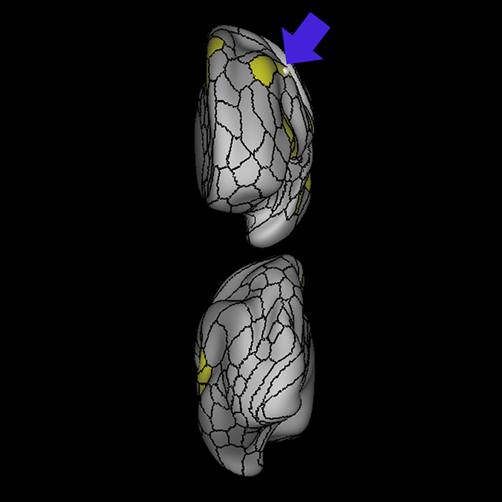
ᐅ SummaryArea SCEF (supplementary and cingulate eye field): part of medial superior frontal gyrus regions. Higher order oculomotor center implicated in appraising all possible oculomotor behaviors to direct primary oculomotor centers in goal- directed behavior. ᐅ Where is it?Area SCEF (supplementary and cingulate eye field) is located in the posterior medial SFG. ᐅ What are its borders?Area SCEF borders area 8BM anteriorly, areas 6ma and SFL superiorly, areas 6mp and 24dd posteriorly, and areas 24dv and p32pr inferiorly. ᐅ What are its functional connections?Area SCEF demonstrates functional connectivity to areas 1, 2, 3a, 3b in the sensory strip, area 4 in the motor strip, areas PEF, FEF, 55b, 6ma, 6mp, 6a, 6r, and 6v in the premotor regions, areas a24prime, p32prime, a32prime, 5mv, and 23c in the middle cingulate regions, areas IFJa, 46, and 9-46d in the lateral frontal lobe areas OP4, OP1, PFcm, 43, FOP1, FOP2, FOP3 FOP4, and FOP5 in the superior insula opercular regions, areas PSL, 52, A4, MI, PoI1 and PoI2 in the lower opercula and Heschl's gyrus regions, area PHT in the temporal lobe, areas AIP, MIP, VIP, LIPd, LIPv, PFop, PF, PFt, IP0, IPS1, 7AL, 7PL, and 7PC, in the lateral parietal lobe, areas 7am and DVT in the medial parietal lobe, area V1, V2, V3, V4 in the medial occipital lobe, areas V3a, V3b, V6, V6a, and V7 in the dorsal visual stream, area FFC of the ventral visual stream, and areas PH, TPOJ2, LO3, MST, and FST of the lateral occipital lobe. ᐅ What are its white matter connections?Area SCEF is structurally connected to the contralateral hemisphere and thalamus. Contralateral connections course through the body of the corpus callosum to end at SCEF, 8BL, SFL and 8BM. Thalamic projections travel through the ventral thalamus to the brainstem (Figure 32). Local short association bundles connect with SF, 8BM, SFL and 8BL. ᐅ What is known about its function?Area SCEF is a higher order oculomotor center implicated in appraising all possible oculomotor behaviors to direct primary oculomotor centers in goal-directed behavior. |
|
A: lateral-medial
B: anterior-posterior
C: superior-inferior
DTI image |
ᐅ SummaryArea 8BM (8B medial): part of medial superior frontal gyrus regions. Involved in maintaining visuospatial information as well as coordinating and coding visuospatial information in terms of oculomotor and other body-centered coordinate systems. ᐅ Where is it?Area 8BM (8B medial) is located in the posterior medial SFG. ᐅ What are its borders?Area 8BM borders area 9m anteriorly and SCEF posteriorly. It borders the following area 24 subdivisions superiorly: a24pr, p24pr, and p24. Its inferior border contains areas d32 and a32pr, and its superior boundary includes 8BL and SFL. ᐅ What are its functional connections?Area 8BM demonstrates functional connectivity to areas i6-8, s6-8, a10p, a9-46v, p9-46v, 8C, 8BL, 8AD, and 8AV in the dorsolateral frontal lobe, areas SFL, a32prime and d32 in the medial frontal lobe, areas IFSa, IFSp, IFJp, 44, 45, a47r, and p47r in the inferior frontal lobe, area 55b in the premotor areas, area AVI in the insula, areas TE1m, TE1p, and STSvp in the temporal lobe, areas LIPv, IP1, IP2, PFm, PGi and PGs in the lateral parietal lobe, and areas 7pm, 31a, and d23ab in the medial parietal lobe . ᐅ What are its white matter connections?Area 8BM is connected to the contralateral hemisphere, frontal aslant tract, inferior front-occipital fasciculus and thalamus. Contralateral connections course through the corpus callosum to end at 8BM and 9m. Frontal aslant tract fibers from 8BM project inferolaterally to end at 44. Thalamic connections run inferior to 8BM and continue in the brainstem. Fibers with the inferior fronto-occipital fasciculus project inferior and posterior through the extreme/external capsule through the temporal lobe to end at parietal and occipital connections 7PC, V1, V2 and V3. Local short association bundles connect with 9m, d32 and SCEF. ᐅ What is known about its function?Area 8Bm is involved in maintaining visuospatial information as well as coordinating and coding visuospatial information in terms of oculomotor and other body-centered coordinate systems. |
|
A: lateral-medial
B: anterior-posterior
C: superior-inferior
DTI image |
ᐅ SummaryArea STSdp (superior temporal sulcus dorsal posterior): part of the temporal lobe regions. Involved in motion processing, audiovisual integration, and facial processing. The posterior half of STSdp (as with STSvp) is strongly activated in the story-math secondary contrast, indicating a role in language comprehension. STSdp responds more strongly than STSvp to primary language tasks and to social cognition and motor tasks. ᐅ Where is it?Area STSdp (superior temporal sulcus dorsal posterior) is found on the posterior half of the lateral face of the STG and the posterior half of the superior bank of the superior temporal sulcus ᐅ What are its borders?Area STSdp borders area STSda anteriorly, STSvp inferiorly, TPOJ1 posteriorly, and A5 superiorly. ᐅ What are its functional connections?Area STSdp demonstrates functional connectivity to areas 9m, 8BL, 44 45, 47L, 47s, IFSp, SFL and 55b in the frontal lobe, areas STV, PSL, A5, and STGa in the insula opercular area, areas STSva, STSvp, STSda, and TGd, in the temporal lobe, TPOJ1 in the lateral occipital lobe, and PGi and 31pd in the parietal lobe ᐅ What are its white matter connections?Area STSdp is structurally connected to the "u" fibers of the occipito-temporal system and the arcuate/SLF. Arcuate/SLF tracts wrap around the Sylvian fissure projecting toward the frontal lobe and turn medially to terminate at 44, FOP4, IFJa, IFJp and IFSp. Local short association fibers include "u" fibers of the occipito-temrporal system that connect to STSda, STSva, STSvp, PSL and P ᐅ What is known about its function?The posterior portion of the STS is primarily involved in motion processing, audiovisual integration, and facial processing. The posterior half of STSdp (like the posterior half of STSvp) is strongly activated in the story-math secondary contrast, indicating a role in language comprehension. STSdp responds more strongly than STSvp to primary language tasks and to social cognition and motor tasks. |
|
A: lateral-medial
B: anterior-posterior
C: superior-inferior
DTI image |

ᐅ SummaryArea STSvp (superior temporal sulcus ventral posterior): part of the temporal lobe regions. Strongly activated by the story-math secondary contrast, indicating a role in language comprehension. STSvp does not respond as strongly as STSva to primary language tasks, and is less active in social cognition and motor tasks. ᐅ Where is it?Area STSvp (superior temporal sulcus ventral posterior) is found on the posterior half of the inferior bank of the superior temporal sulcus ᐅ What are its borders?Area STSvp borders area STSva anteriorly, TE1m and TE1p inferiorly, TPOJ1 and PHT posteriorly, and STSdp superiorly ᐅ What are its functional connections?Area STSvp demonstrates functional connectivity to 8AV, 8BL, 8BM, 8C, 9a, 9p, 9m, 44, 45, 47s, 47L a47r, IFSp, d32, 10v, SFL and 55b in the frontal lobe, area PSL in the insula opercular area, areas STSva, STSda, STSdp, TGd, TE1a, TE1m, TE1p, and TE2a in the temporal lobe, and areas PGs, PGi, 7m, POS1, d23ab, 31pv, and 31pd in the parietal lobe ᐅ What are its white matter connections?Area STSvp is structurally connected to “u” fibers of the occipito-temporal system and the arcuate/SLF. Arcuate/SLF tracts wrap around the Sylvian fissure projecting toward the frontal lobe to turn medially and terminate at 6r, IFJp, IFJa, FOP2, FOP3, FOP4 and 44. There are posterior projections from the arcuate/SLF as it wraps around the Sylvian fissure to terminate at the inferior parietal lobule at PF, PFm, PSL, PGi and STV. Local short association fibers include “u” fibers of the occipito-temrporal system that connect to STSdp, STSva, TE1p, TE1m, PF, PFm, PSL, PGi and STV ᐅ What is known about its function?The posterior half of STSvp (like the posterior half of STSdp) is strongly activated by the story-math secondary contrast, indicating a role in language comprehension. STSvp does not respond as strongly as STSva to primary language tasks, and is less active in social cognition and motor tasks |
|
A: lateral-medial
B: anterior-posterior
C: superior-inferior
DTI image |
ᐅ SummaryArea AIP (anterior intraparietal): part of the lateral parietal lobe regions. Involved in grasping activity as well as object recognition. Neurons in this part of the cortex are oriented for grip and hand shape, and the inferior temporal cortex provides input related to object information. Receives input from the ventral and dorsolateral visual streams. Plays a role in shaping the hand for grasping activity. Beyond grasping action, is involved in tactile shape-processing and understanding orientation in space. ᐅ Where is it?Area AIP (Anterior intraparietal) is found on the superior bank of the intraparietal sulcus at its most anterior aspect. It extends onto the superior surface of the adjacent superior parietal lobule, and its anterior tip lies in the bank of the postcentral sulcus. ᐅ What are its borders?Area AIP borders Area 2 anteriorly and PFt anteroinferiorly. Its anterosuperior border is area 7PC, and its inferior border is with IP2 across the intraparietal sulcus. LIPv and LIPd make up its posterior boundaries. ᐅ What are its functional connections?Area AIP demonstrates functional connectivity to area 2 in the sensory strip, areas SCEF, FEF, PEF 6a, 6r, and 6ma in the premotor regions, areas IFSa, IFJp, 46, and p9-46v in the lateral frontal lobe, areas 5mv, and 23c in the medial frontal lobe, areas PoI2, FOP2, FOP4, and OP4 in the insula opercular regions, areas PHA3, PHT and TE2p, in the temporal lobe, areas 7PC, 7PL, 7AL, VIP, MIP, LIPv, LIPd, PFop, PFt, PGp, IP2, IP1, IP0, and IPS1 in the lateral parietal lobe, areas DVT, 7AM and 7pm in the medial parietal lobe, area V2 in the medial occipital lobe, area FFC in the ventral visual stream areas, and areas V3CD, PH, TPOJ2, and FST in the lateral occipital lobe. ᐅ What are its white matter connections?Area AIP is structurally connected to the pars opercularis and local parcellations. Connections from AIP to the pars opercularis travel anteroinferiorly to end at 43 and 6r. Local short association bundles connect with 2, PFt, PFcm, PF, IP2 and 7PC. ᐅ What is known about its function?Area AIP is involved in grasping activity as well as object recognition. Neurons in this part of the cortex are oriented for grip and hand shape, and the inferior temporal cortex provides input related to object information. AIP also receives input from the ventral and dorsolateral visual streams. This region also plays a role in shaping the hand for grasping activity. Beyond grasping action, AIP is involved in tactile shape-processing and understanding orientation in space. |
|
A: lateral-medial
B: anterior-posterior
C: superior-inferior
DTI image |
ᐅ SummaryArea PFm (parietal area F, part m): part of the lateral parietal lobe regions. Shows activation in nonspatial attention tasks, decision making when switching choices, rule change during visually guided attention, and reorientation. Also provide syntactical components to language processing, plays a role in attentional processing, and is activated in working memory, motor cue, and risk-related tasks. ᐅ Where is it?Area PFm (parietal area F, part m) is found on the anterior superior surface of the angular gyrus, and straddles the sulcus to lie on the posterior superior bank of the supramarginal gyrus. ᐅ What are its borders?Area PFm borders IP2 and IP1 superiorly, PF anteriorly, PSL and STV inferiorly, and PGI and PGS posteriorly. ᐅ What are its functional connections?Area PFm demonstrates functional connectivity to areas 8AV, 8AD, 8BL, 8C, s6-8, i6-8, a47r, p47r, a10p, p10p, 9a, a9-46v, p9-46v, and area 44 in the lateral frontal lobe, area d32 in the medial frontal lobe, area AVI in the insula, areas STSvp, TE1m, TE1p, and TE2a, in the temporal lobe, areas PGs, PGi, IP2, and IP1 in the lateral parietal lobe, and areas 7m, 7pm, POS2, 31a, 31pv, d23ab, 23d and RSC in the medial parietal lobe. ᐅ What are its white matter connections?Area PFm is structurally connected to the arcuate/SLF. Arcuate/SLF connections course anteriorly from PFm to 8C and 8BM, and inferiorly to middle temporal gyrus parcellations TE1a, TE1m, TE1p, STSva, STSvp and PHT. Local short association bundles connect with AIP, 7PC, IP1, IP2, LIPd, LIPv, PGi, PGs, 2 and 1 ᐅ What is known about its function?PFm shows activation in non-spatial attention tasks, decision making when switching choices, rule change during visually-guided attention, and reorientation. Intermediate regions of the inferior parietal lobule also provide syntactical components to language processing. This area also plays a role in attentional processing, and is activated in working memory, motor cue, and risk-related tasks. |
|
A: lateral-medial
B: anterior-posterior
C: superior-inferior
DTI image |
ᐅ SummaryArea TE1p (temporal area 1 posterior): part of the temporal lobe regions. Appears primarily related to visual pathways. TE1p, like TE1m, shows greater activation in the visual working memory secondary contrast compared to area TE1a. Relative to TE1m, TE1p is more deactivated during language tasks and more activated during facial recognition tasks. ᐅ Where is it?Area TE1p (TE 1 posterior) is found on the posterior most portions of the middle an inferior temporal gyri and the intervening inferior temporal sulcus. It spills onto the basal face of the temporal lobe and extends up to the occipitotemporal sulcus. ᐅ What are its borders?Area TE1p borders TE1m and TE2a anteriorly. Its posterior border is made up of PHT on the lateral surface and PH on the basal surface. TE2p forms its inferobasal edge and STSvp forms its superior edge. ᐅ What are its functional connections?Area TE1p demonstrates functional connectivity to 33prime, 8AV, 8AD, 8BM, 8C, IFSa, IFSp, IFJp a47r, p47r, 47m, a9-46v, p9-46v, i6-8 and s6-8 in the frontal lobe, areas STSvp, PHT, TE1m, and TE2a in the temporal lobe, and areas PGs, PGi, PFm, IP2, IP1, IP0, 7pm, 7m, d23ab, and 31a in the parietal lobe. ᐅ What are its white matter connections?Area TE1p is structurally connected to the arcuate/SLF and "u" fibers of the occipito-temporal system. Arcuate/SLF tracts wrap around the Sylvian fissure projecting toward the frontal lobe to turn medially and end at 45. There are abundant posterior projections from the arcuate/SLF that terminate at the inferior parietal lobule at STV, PFm, PSL, PGi, TPOJ1, TPOJ2 and STV. Local short association fibers include "u" fibers of the occipito-temrporal system that connect to TE2a and PeEc. ᐅ What is known about its function?The function of area TE1p appears primarily related to visual pathways. TE1p, like TE1m, shows greater activation in the visual working memory secondary contrast compared to area TE1a. Relative to TE1m, TE1p is more deactivated during language tasks and more activated during facial recognition tasks. |
|
A: lateral-medial
B: anterior-posterior
C: superior-inferior
DTI image |
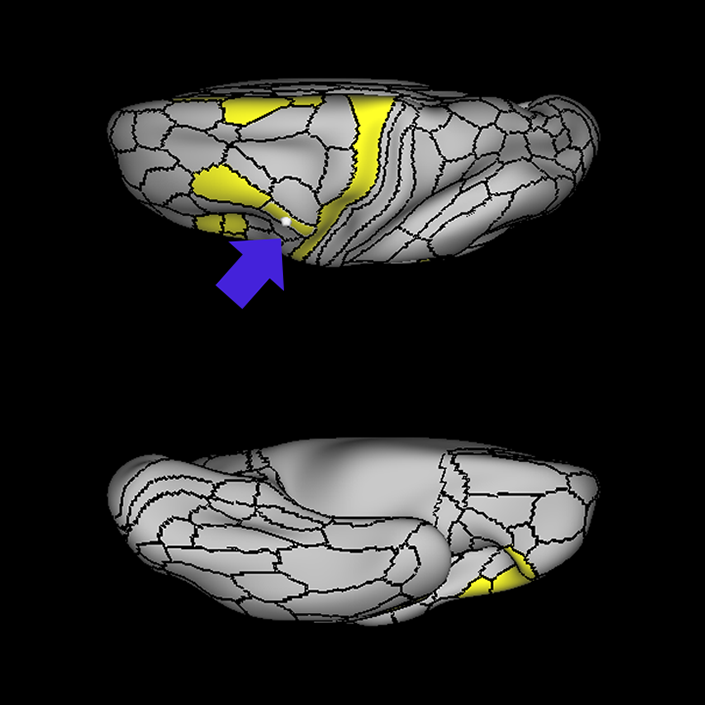
ᐅ SummaryArea PHT (parahippocampal temporal): part of the temporal lobe regions. Involved in processes related to the controlled retrieval of conceptual knowledge, while the anterior gyrus is involved in the automatic retrieval of specific semantic information. In contrast to the other areas of the lateral temporal cortex and temporal pole, that are all strongly associated with the task negative network, PHT is strongly associated with the task positive network. In addition, PHT (like TE1p anteriorly) is deactivated during language recognition tasks. ᐅ Where is it?Area PHT is found on the anterior portions of the subcentral gyrus (where the precentral and postcentral gyri meet just below the central sulcus). It involves the lateral surface of that operculum as well as the inferior surface which faces the Sylvian Fissure. ᐅ What are its borders?Area PHT borders area 6r anteriorly and OP4 posteriorly. Its superior border includes area 6v, as well as areas 4 and 3a. Its inferior borders include FOP1 and FOP2. ᐅ What are its functional connections?Area PHT demonstrates functional connectivity to areas IFSa, IFJa, IFJp, 6a,6ma, 6r, 46, 9-46d, p9-46v, p47r, FEF, PEF, SCEF, a24prime, p24prime, p32prime, 33prime, 23c, and 5mv in the frontal lobe, areas FOP1, FOP3, FOP4, FOP5, 43, PFcm, 52, MI, PoI1, and PoI2 in the insula opercular area, areas TE1p, TE2p and PHA3 in the temporal lobe, areas AIP, MIP, VIP, LIPv, LIPd, IPS1, IP0, IP1, IP2, PF, PFop, PFt, PGp, 7PC, 7pm, 7AL, 7PL, PCV and DVT in the parietal lobe, and areas V1, V2, FST, PH, TPOJ2 in the occipital lobe. ᐅ What are its white matter connections?Area PHT is structurally connected to the arcuate/SLF. Arcuate/SLF tracts wrap around the Sylvian fissure projecting toward the frontal lobe and turn medially to end at 44, IFJa, IFJp and IFSp. There are abundant posterior projections from the arcuate/SLF that terminate at the inferior parietal lobule at PGs, STV, PFm, PGi, TPOJ1 and TPOJ2. ᐅ What is known about its function?Area PHT lies in the posterior MTG leading into the angular gyrus. The posterior MTG is involved in processes related to the controlled retrieval of conceptual knowledge, while the anterior gyrus is involved in the automatic retrieval of specific semantic information. In contrast to the other parcellations of the lateral temporal cortex and temporal pole (TE1p, TE1m, TE1a, TE2p, TE2a, TGv, TGd, and TF) which are all strongly associated with the task negative network, PHT is strongly associated with the task positive network. In addition, PHT (like TE1p anteriorly) is deactivated during language recognition tasks. |
|
A: lateral-medial
B: anterior-posterior
C: superior-inferior
DTI image |
ᐅ SummaryArea PBelt (parabelt complex): part of parietal apex regions and superior insula opercular regions. Parcellated from the auditory cortex. ᐅ Where is it?Area PBelt (Parabelt complex) is located on the superior surface of the inferior portion of the supramargina gyrus. It lies in the small region between the lateral edge of Heschl's gyrus and the opercular cleft of the inferior SMG. ᐅ What are its borders?Area PBelt borders Lbelt and Mbelt medially and A4 laterally. Its deep surface borders with RI ᐅ What are its functional connections?Area PBelt demonstrates functional connectivity to areas 1, 2, 3a, and 3b in the sensory strip, area 4 in the motor strip, area 24dd in the paracingulate areas, areas FEF, 6d, 6v, and 6mp in the premotor areas, areas 43, PFcm, OP1, OP2-3, and OP4 in the superior opercular region, areas A1, A4, A5 Mbelt, LBelt, PoI1, PoI2, STV, Ta2, PI, RI, and 52 in the inferior insula opercular region, areas PFop and 7PC in the parietal lobe, areas V1, V2, V3, and V4 in the medial occipital lobe, areas V6, V67a, V7, V3a, and V3b in the dorsal visual stream, areas V8, FFC, Pit and VVC in the ventral visual stream, and areas LO2, LO3, V3cd, FST, MT, MST, V4t, TPOJ1, and TPOJ2 in the lateral occipital lobe. ᐅ What are its white matter connections?Area PBelt is structurally connected to the middle longitudinal fasciculus and arcuate/SLF. Arcuate/SLF fibers wrap around the termination of the sylvian fissure to end at the inferior frontal gyrus at parcellation 45 and FOP5. Fibers from the middle longitudinal fasciculus project posterior just lateral to the lateral ventricle to occipital and parietal lobes to end at parcellations MIP, LIPv and IP1. Local short association bundles connect with A1, LBelt, MBelt, PFcm, PSL, A4, A5, TPOJ1. ᐅ What is known about its function?Area PBelt is a newly described area of the brain parcellated from the auditory cortex. Area PBelt was differentiated from areas A4 and LBelt based on differences in activity on fMRI during arithmetic, auditory story, and motor cue tasks. |
|
A: lateral-medial
B: anterior-posterior
C: superior-inferior
DTI image |