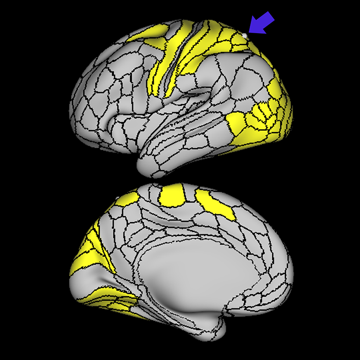
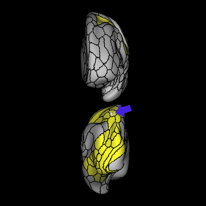
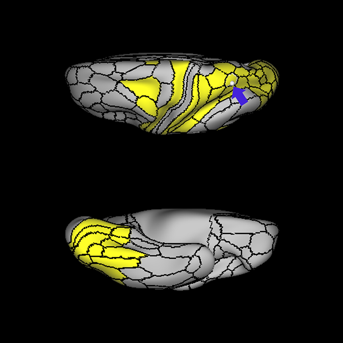
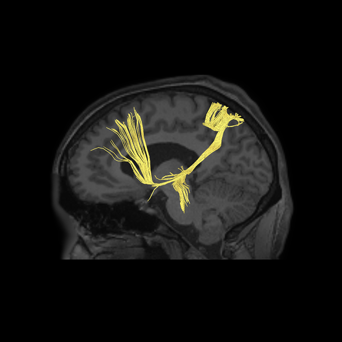
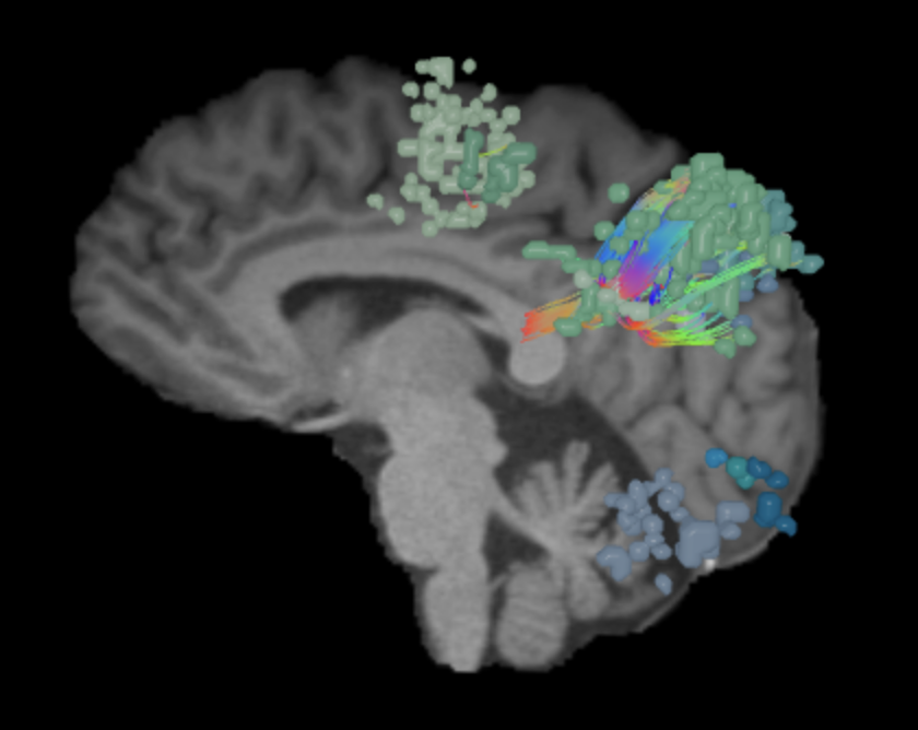
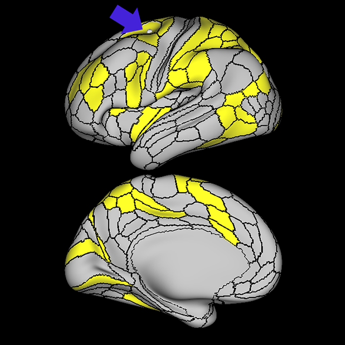
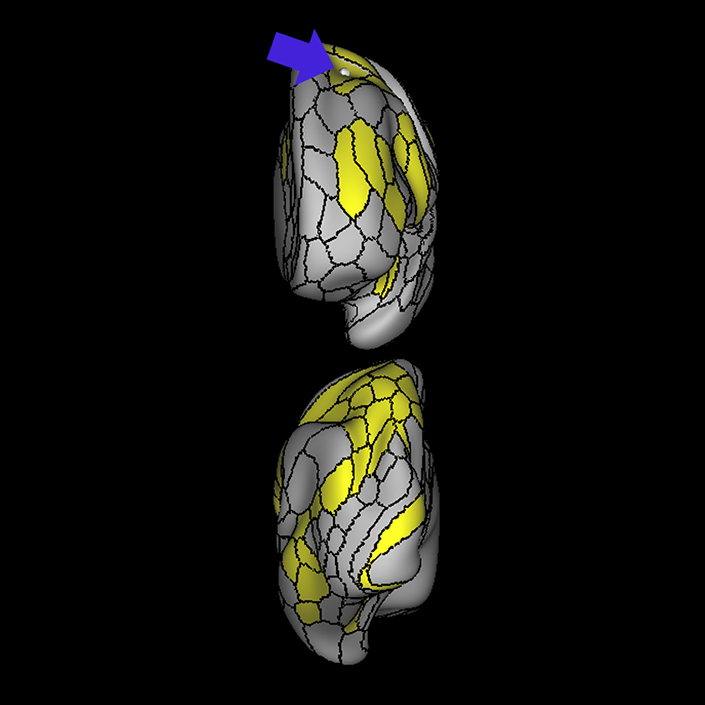
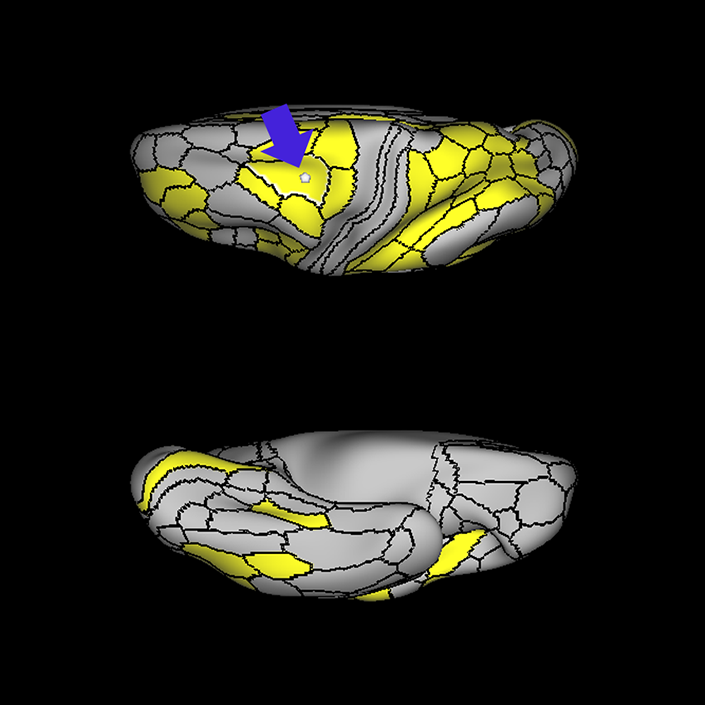
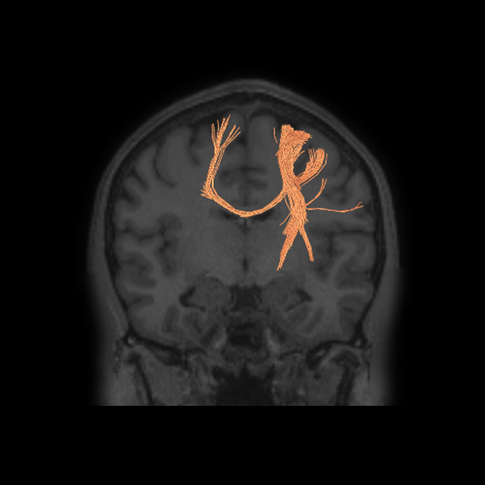
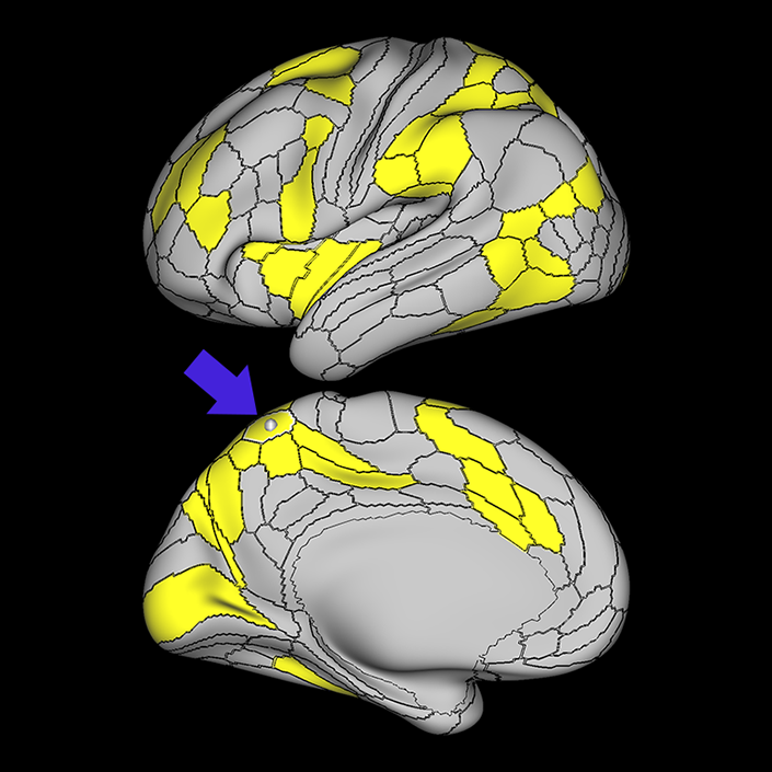
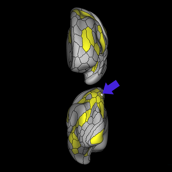
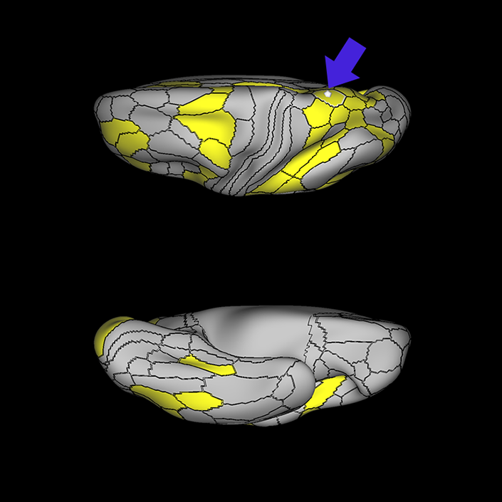
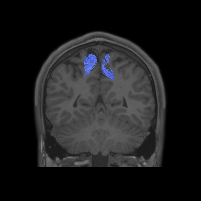
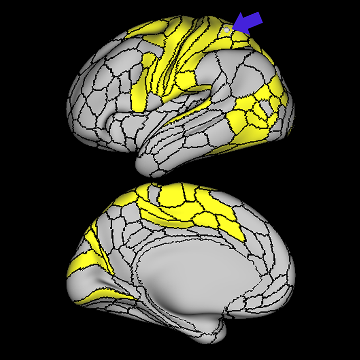
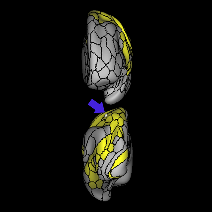
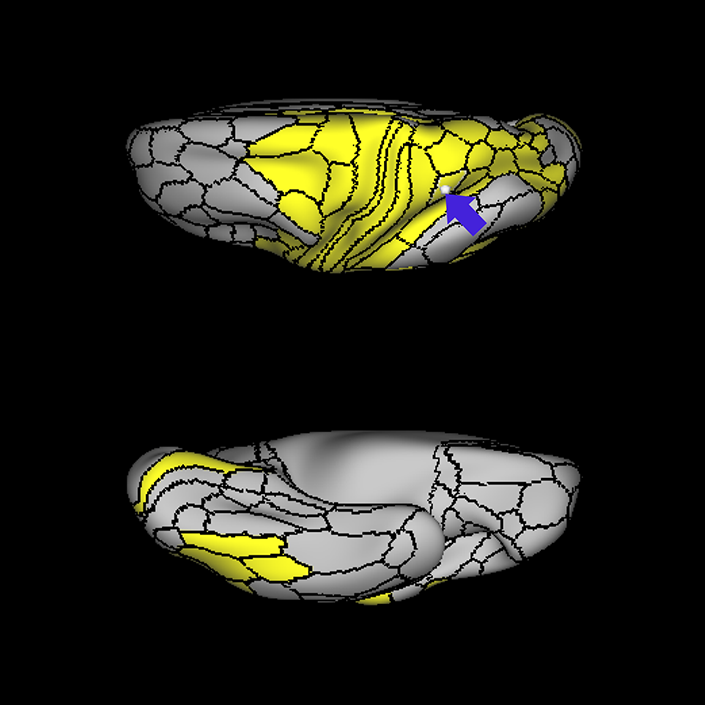
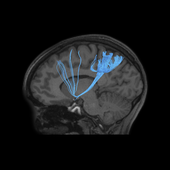
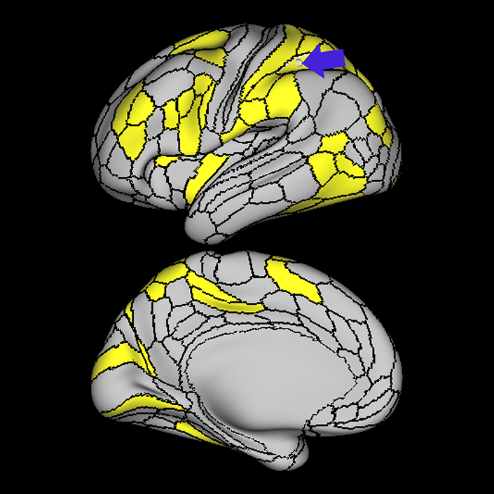
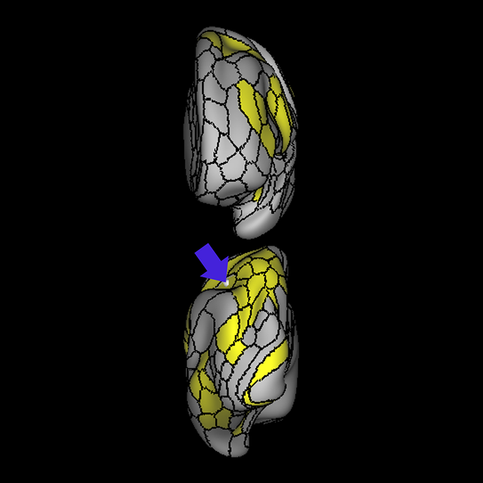
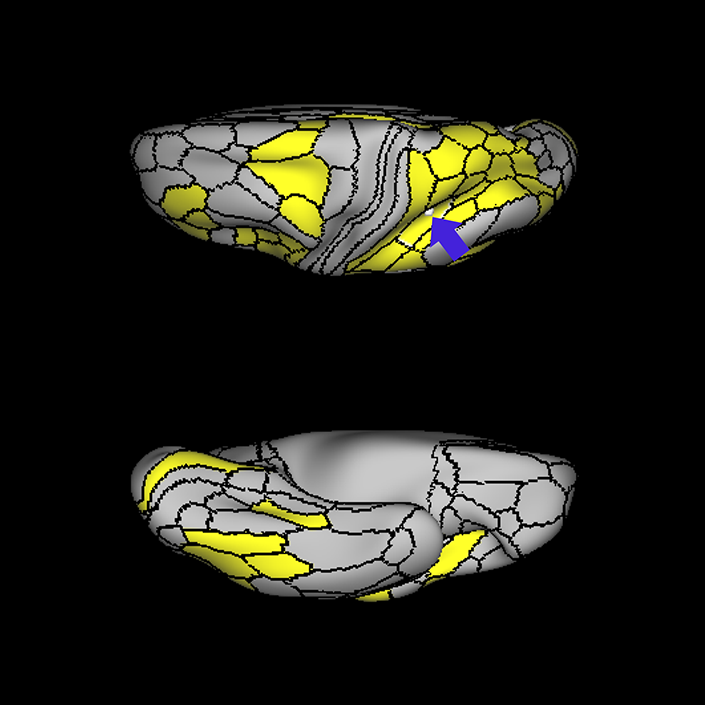
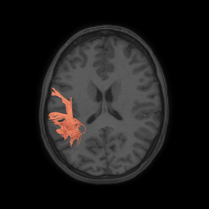
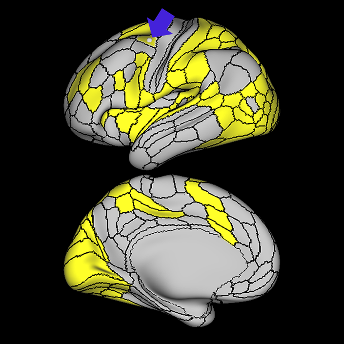
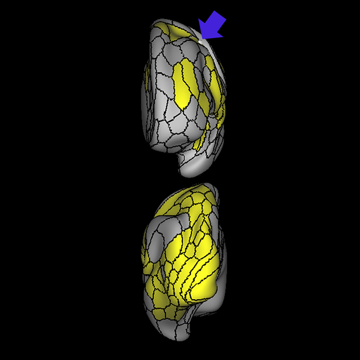
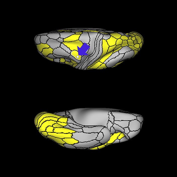
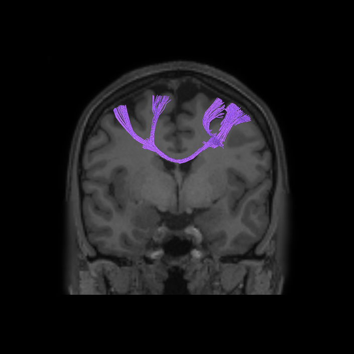
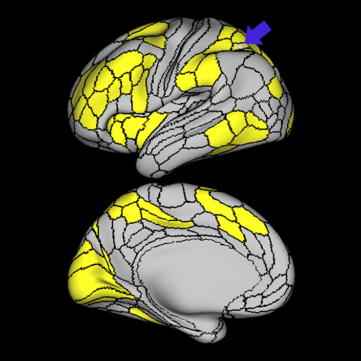
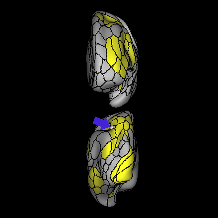
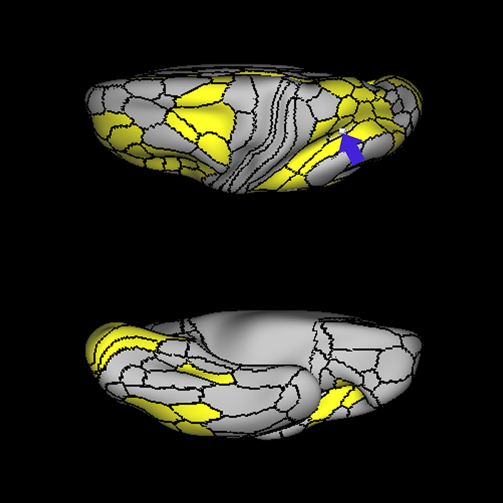
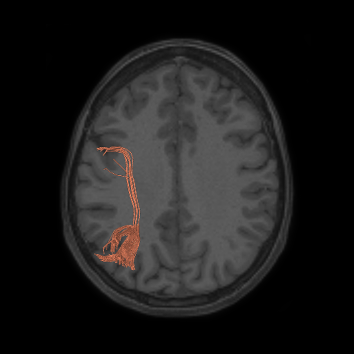
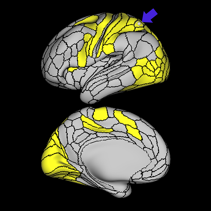
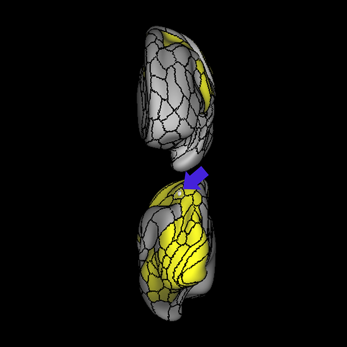
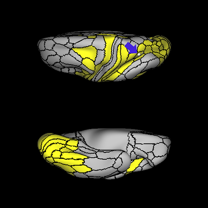
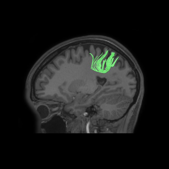
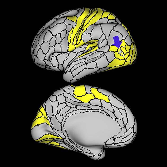
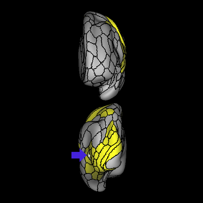
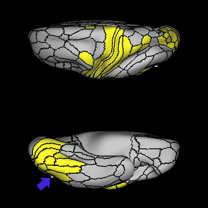
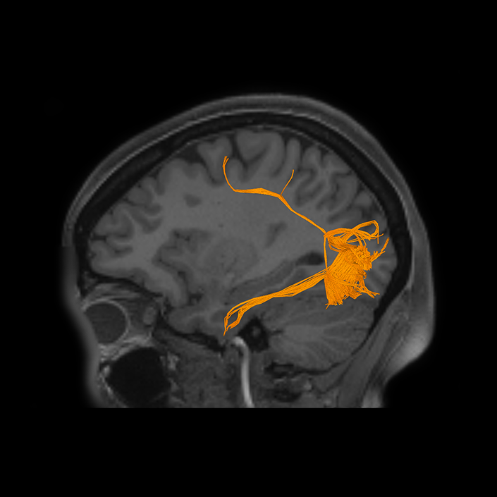
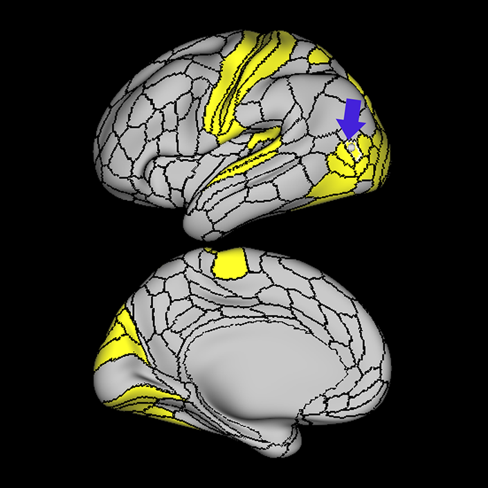
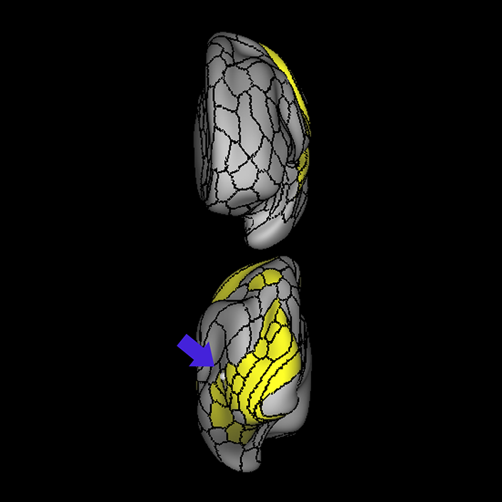
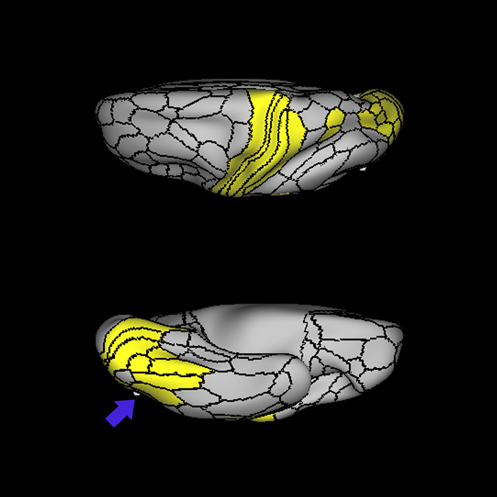
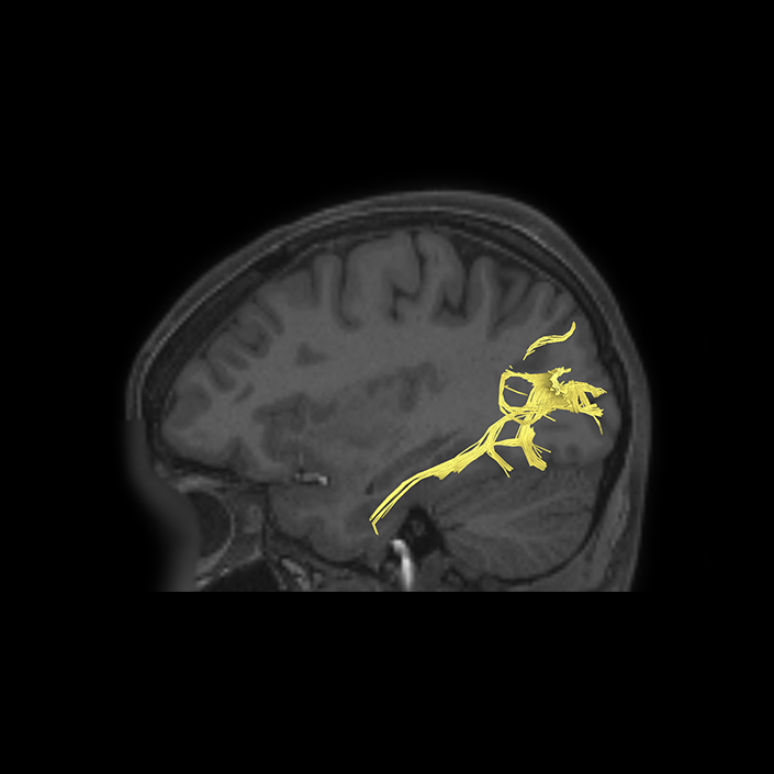
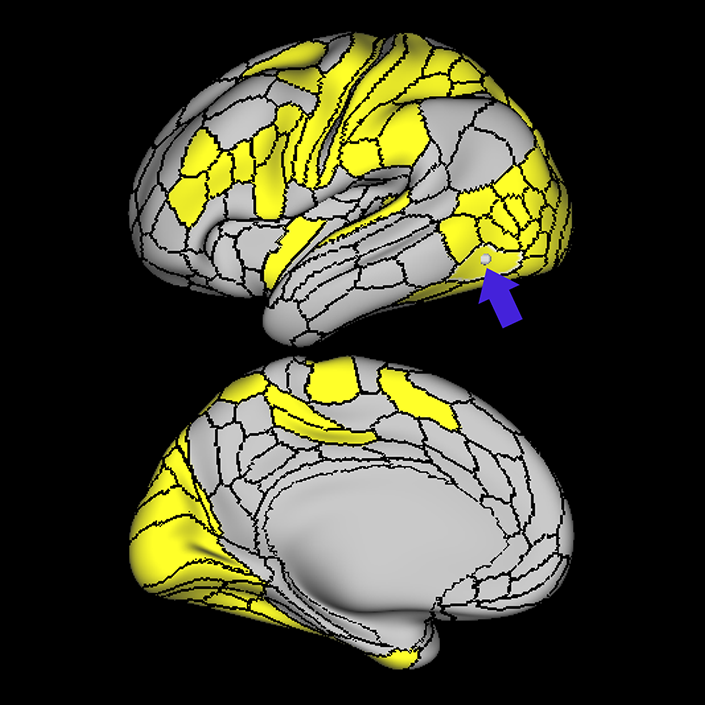
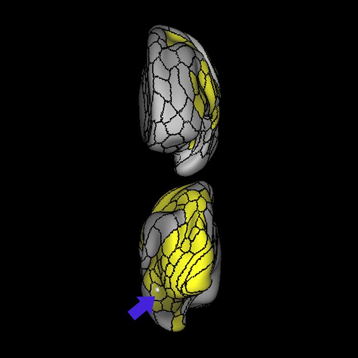
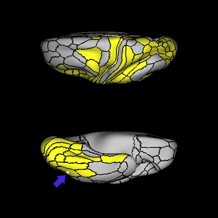
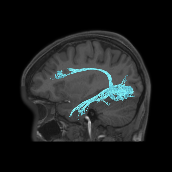
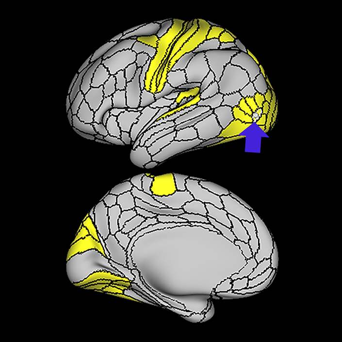
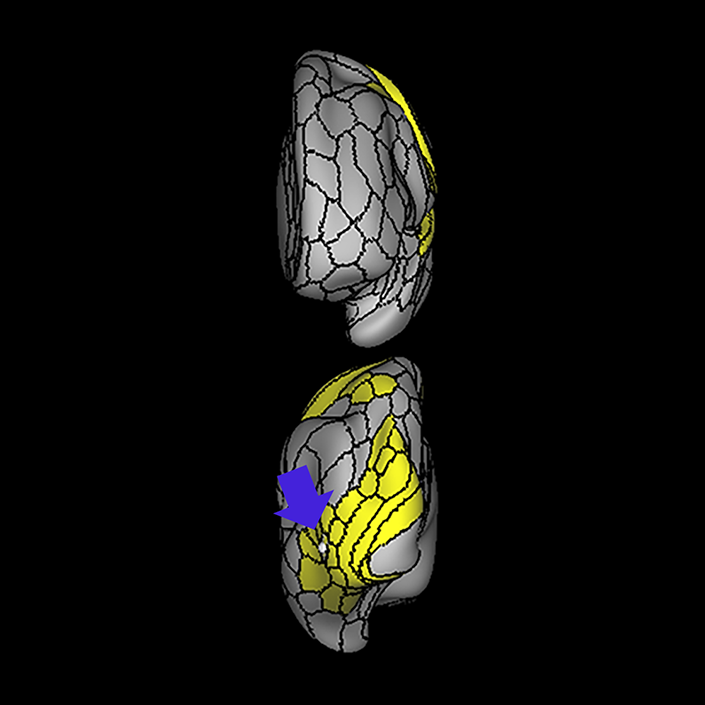
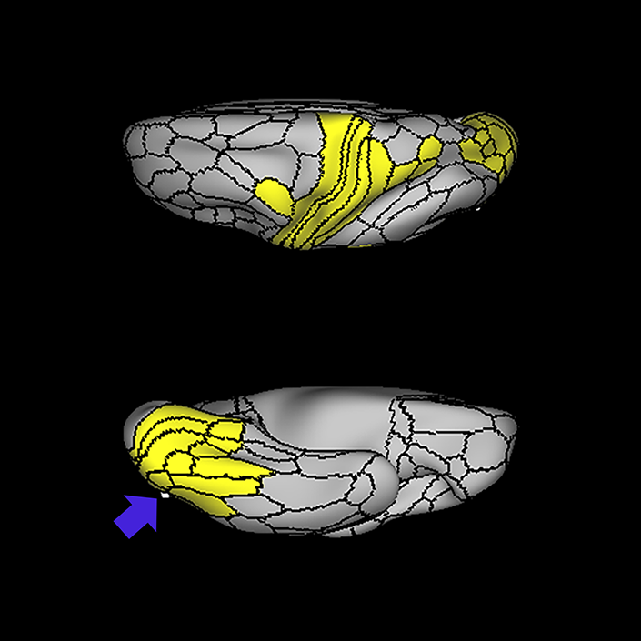
ᐅ SummaryArea 6a (6 anterior): part of the premotor areas. While the precise function of this area is unknown, the functions of the premotor cortex are well characterized in the literature. 6a shows greater activation when solving math problems, in social interaction settings, and when performing object feature comparison tasks. ᐅ Where is it?Area 6a (6 anterior) makes up the posterior superior most bank of the superior frontal sulcus and the adjacent portions of the superior frontal gyrus, principally forming this bank just as the sulcus forms the right angle with the precentral sulcus. ᐅ What are its borders?Area 6a borders areas 6d and 6mp posteriorly, and area 6ma medially. Its anterior border is made by s6-8, area 8AD, and i6-8. FEF is its inferior neighbor. ᐅ What are its functional connections?Area 6a demonstrates functional connectivity to area 2 in the sensory strip, areas SCEF, PEF, FEF, 6ma, 6mp, 6d, and 6v in the premotor regions, areas a24prime, p32prime, 5mv, and 23c in the middle cingulate regions, areas IFSa, IFJa, i6-8, 46, p9-46v and 9-46d in the lateral frontal lobe areas OP4, PFcm, FOP4, and FOP2 in the superior insula opercular regions, areas PoI1 and PoI2 in the lower opercula and Heschl's gyrus regions, areas TE2p, PHA3 and PHT in the temporal lobe, areas AIP, MIP, VIP, LIPd, LIPv, PFop, PF, PFt, PGp IP2, IP1, IP0, IPS1, 7AL,7PL, and 7PC, in the lateral parietal lobe, areas 7pm, 7am, DVT, and PCV in the medial parietal lobe, area V2 in the medial occipital lobe, and areas PH, TPOJ2, TPOJ3, and FST of the lateral occipital lobe. ᐅ What are its white matter connections?Area 6a is structurally connected to the pyramidal tracts and the parietal lobe. Connections to pyramidal tracts descend through the posterior limb of the internal capsule and cerebral peduncle to the brainstem. Parietal projections are portions of the SLF and connect with 3a, 3b, 7PC and 7AL. Local short association fibers connect with FEF, i6- 8, 55b, 8Av, 46 and 6r. ᐅ What is known about its function?Areas 6a and 6d are newly described subdivisions of the premotor cortex. While the precise function of these areas is unknown, the functions of the premotor cortex are well characterized in the literature. First, the premotor cortex is functionally divided into ventral and dorsal aspects. The dorsal premotor cortex is involved in associating informational cues with a particular body movement. These cues could be learned and arbitrary in nature or they can be based on other forms of somatosensation, such as visual or auditory sensation. The ventral premotor area is involved in hand movement manipulation of objects, e.g. when grasping or lifting. The ventral premotor area is also involved in more complex cortical functions such as when individuals learn actions or movements while observing others performing a task. Overall, the premotor cortex has significant function in the preparation of voluntary movements. Regarding area 6a more specifically, this region was distinguished from adjacent areas of the cortex based on differences in myelin thickness and functional activity.9 Compared to area s6-8, area 6a shows greater activation when solving math problems, in social interaction settings, and when performing object feature comparison tasks. Compared to area i6-8, area 6a shows greater activation in social interaction settings and relative deactivation in emotion identification and object feature comparison. Compared to area FEF, area 6a shows less activation in both gambling and object feature comparison. |
|
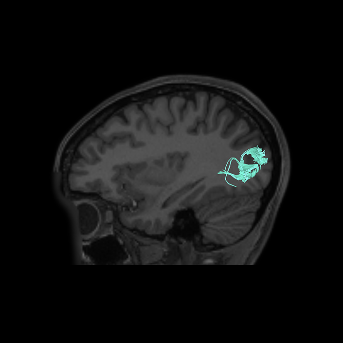
A: lateral-medial
B: anterior-posterior
C: superior-inferior
DTI image |
ᐅ SummaryArea 7Am (7 anterior-medial): part of the lateral parietal lobe regions. Involved in several types of information processing including space, vision shape and motion, working memory, and execution. The anterior portion of 7Am is involved in self-centered mental imagery and attentional processes. Relative to its posterolateral neighbor 7PL, area 7Am is less activated during working memory and auditory story tasks. Relative to its posteromedial neighbor 7Pm, area 7Am is less activated during working memory and shape recognition tasks. ᐅ Where is it?Area 7AM (7 anterior-medial) is found on the anterosuperior surface of the medial face of the superior parietal lobule. ᐅ What are its borders?Area 7AM borders the area 7AL superior-laterally, VIP posterolaterally, and areas 7PL and 7PM posteriorly. On its interhemispheric face, its anterior border is made up of areas 5L, and 5MV. Its inferior border is made up of PCV (precuneus visual area), and its posterior border is made up of area 7PM. ᐅ What are its functional connections?Area 7AM demonstrates functional connectivity to areas SCEF, FEF, PEF, 6ma, 6a, and 6r in the premotor regions, areas IFSa, 46, and 9-46d areas in the lateral frontal lobe, areas a24prime, a32prime, p32prime, 5mv, and 23c in the medial frontal lobe, areas MI, PoI1, PoI2, PFcm and FOP4 in the insula opercular regions, areas PHA3, PHT, and TE2p, in the temporal lobe, areas 7PC, 7AL, 7PL, AIP, VIP, MIP, LIPd, PFop, PFt, PF, PGp, IP2, and IP0 in the lateral parietal lobe, areas 7pm, PCV, POS2, and DVT in the medial parietal lobe, area V1 in the medial occipital lobe, areas V6 in the dorsal visual stream areas, area FFC in the ventral visual stream areas, and areas PH, TPOJ2, TPOJ3, and FST in the lateral occipital lobe. ᐅ What are its white matter connections?Area 7AM is structurally connected to the contralateral hemisphere and thalamus. Some individuals have IFOF connections but these tracts are inconsistent. Contralateral connections course through the corpus callosum to end at 7AM and 7Pm. Thalamic connections project inferior through the posterolateral thalamus to the brainstem and superior colliculus. Local association bundles connect with VIP and 7PL. ᐅ What is known about its function?Area 7AM is involved in several types of information processing including space, vision shape and motion, working memory, and execution. The anterior portion of 7AM is involved in self-centered mental imagery and attentional processes. Relative to its posterolateral neighbor 7PL, area 7AM is less activated during working memory and auditory story tasks. Relative to its posteromedial neighbor 7PM, area 7AM is less activated during working memory and shape recognition tasks. |
|
A: lateral-medial
B: anterior-posterior
C: superior-inferior
DTI image |
ᐅ SummaryArea 7PC (7 postcentral): part of the lateral parietal lobe regions. Involved in vision motion, observation, space, and execution. The left hemispheric portion of region 7PC is associated with imagination, and the right region is associated with vision shape, language comprehension, sexuality, and working memory. This region is involved in visual and somatosensory stimulation, and shows strong connection to somatosensory areas. ᐅ Where is it?Area 7PC (7 postcentral) is found in the anterior, inferior superior parietal lobule. It extends into the adjacent posterior bank of the postcentral sulcus. ᐅ What are its borders?Area 7PC borders Area 2 anteriorly and AIP inferiorly. Its posterior border is made up of LIPv and VIP. Area 7AL is its medial (superior) neighbor. ᐅ What are its functional connections?Area 7PC demonstrates functional connectivity to areas 1, 2, 3a, and 3b in the sensory strip, area 4 in the motor strip, areas SCEF, FEF, PEF 6ma, 6mp, 6a, 6d, 6r, and 6v in the premotor regions, areas 24dd, 24dv, p32prime, 5L, 5mv, and 23c in the medial frontal lobe, areas A4, PBelt, PFcm, FOP2, OP4, and OP1 in the insula opercular regions, areas PHT and TE2p, in the temporal lobe, areas 7PL, AIP, VIP, MIP, LIPv, LIPd, PFop, PFt, PGp, IP0, and IPS1 in the lateral parietal lobe, areas 7AM and DVT in the medial parietal lobe, area V2 in the medial occipital lobe, areas V3b, V6a, and V6 in the dorsal visual stream areas, area FFC in the ventral visual stream areas, and areas V3CD, V4t, PH, LO3, TPOJ2, TPOJ3, MST, and FST in the lateral occipital lobe. ᐅ What are its white matter connections?Area 7PC is structurally connected to local parcellations and the IFOF. Connections from the IFOF course through the posterior temporal gyrus and extreme/external capsule to the frontal lobe to 8BL, 6a, 6ma and SFL. The majority of individuals have IFOF projections but this tract is not present in everyone. Local short association bundles are abundant and connect with LIPd, LIPv, MIP, 1 and 2. Whitematter connections from 7PC in the right hemisphere have more consistent connections with the motor and somatosensory cortex. ᐅ What is known about its function?Area 7PC is involved in vision motion, observation, space, and execution. The left hemispheric portion of region 7PC is associated with imagination, and the right region is associated with vision shape, language comprehension, sexuality, and working memory. This region is involved in visual and somatosensory stimulation, and shows strong connection to somatosensory areas. |
|
A: lateral-medial
B: anterior-posterior
C: superior-inferior
DTI image |
ᐅ SummaryArea AIP (anterior intraparietal): part of the lateral parietal lobe regions. Involved in grasping activity as well as object recognition. Neurons in this part of the cortex are oriented for grip and hand shape, and the inferior temporal cortex provides input related to object information. Receives input from the ventral and dorsolateral visual streams. Plays a role in shaping the hand for grasping activity. Beyond grasping action, is involved in tactile shape-processing and understanding orientation in space. ᐅ Where is it?Area AIP (Anterior intraparietal) is found on the superior bank of the intraparietal sulcus at its most anterior aspect. It extends onto the superior surface of the adjacent superior parietal lobule, and its anterior tip lies in the bank of the postcentral sulcus. ᐅ What are its borders?Area AIP borders Area 2 anteriorly and PFt anteroinferiorly. Its anterosuperior border is area 7PC, and its inferior border is with IP2 across the intraparietal sulcus. LIPv and LIPd make up its posterior boundaries. ᐅ What are its functional connections?Area AIP demonstrates functional connectivity to area 2 in the sensory strip, areas SCEF, FEF, PEF 6a, 6r, and 6ma in the premotor regions, areas IFSa, IFJp, 46, and p9-46v in the lateral frontal lobe, areas 5mv, and 23c in the medial frontal lobe, areas PoI2, FOP2, FOP4, and OP4 in the insula opercular regions, areas PHA3, PHT and TE2p, in the temporal lobe, areas 7PC, 7PL, 7AL, VIP, MIP, LIPv, LIPd, PFop, PFt, PGp, IP2, IP1, IP0, and IPS1 in the lateral parietal lobe, areas DVT, 7AM and 7pm in the medial parietal lobe, area V2 in the medial occipital lobe, area FFC in the ventral visual stream areas, and areas V3CD, PH, TPOJ2, and FST in the lateral occipital lobe. ᐅ What are its white matter connections?Area AIP is structurally connected to the pars opercularis and local parcellations. Connections from AIP to the pars opercularis travel anteroinferiorly to end at 43 and 6r. Local short association bundles connect with 2, PFt, PFcm, PF, IP2 and 7PC. ᐅ What is known about its function?Area AIP is involved in grasping activity as well as object recognition. Neurons in this part of the cortex are oriented for grip and hand shape, and the inferior temporal cortex provides input related to object information. AIP also receives input from the ventral and dorsolateral visual streams. This region also plays a role in shaping the hand for grasping activity. Beyond grasping action, AIP is involved in tactile shape-processing and understanding orientation in space. |
|
A: lateral-medial
B: anterior-posterior
C: superior-inferior
DTI image |
ᐅ SummaryArea FEF: part of the premotor areas. Known to be involved in rapid eye movements between fixed points, also known as intentional saccadic movements. Area FEF has also been implicated in smooth eye movements that allow the eyes to follow a moving target, also known as smooth pursuit eye movements. Together, these movements help the FEF to create a salience map for visual attention. ᐅ Where is it?Area FEF is located on the anterior half of the precental gyrus, approximately half way down its length along the convexity, just inferior to the junction point of the precental and superior frontal sulci. It also forms the adjacent floor of the precentral sulci and straddles slightly onto the posterior edge of the middle frontal gyrus. ᐅ What are its borders?Area FEF borders areas 6a and 6d superiorly and area 55b inferiorly. Area 4 is its posterior border and area i6-8 forms its anterior border on the middle frontal gyrus. ᐅ What are its functional connections?Area FEF demonstrates functional connectivity to area 2 in the sensory strip, areas SCEF, PEF, 6r, and 6v in the premotor regions, areas a24prime, p32prime, 5mv, and 23c in the middle cingulate regions, areas IFSa, IFJa, 46, and 9-46d in the lateral frontal lobe, areas 43, OP4, PFcm, FOP1, FOP3, FOP4, and FOP5 in the superior insula opercular regions, areas STV, LBelt, PBelt, A4, MI, 52, RI, PoI1 and PoI2 in the lower opercula and Heschl's gyrus regions, areas TE2p and PHT in the temporal lobe, areas AIP, MIP, VIP, LIPd, LIPv, PFop, PF, PFt, PGp, IP0, IPS1, 7AL,7PL, and 7PC, in the lateral parietal lobe, areas 7am, DVT, and PCV in the medial parietal lobe, areas V1, V2, V3 and V4 in the medial occipital lobe, areas V3a, V3b, V6, V6a, and V7 of the dorsal visual stream, areas V8 PIT, FFC, VVC, VMV1, VMV2, and VMV3 of the ventral visual stream, and areas V3cd, LO1, LO2, LO3, PH, TPOJ1, TPOJ2, TPOJ3, V4t, MST, and FST of the lateral occipital lobe. ᐅ What are its white matter connections?Area FEF is structurally connected to the contralateral hemisphere and superior longitudinal fasciculus. Contralateral connections course through the body of the corpus callosum to i6-8 and SFL. Connections with the superior longitudinal fasciculus connect FEF to the intraparietal sulcus and the inferior parietal lobe terminating at IP1, IP2 and PGs. Local short association fibers connect with 6d, 55b, i6-8, 8Av, 6a and PEF. ᐅ What is known about its function?Area FEF is known to be involved in rapid eye movements between fixed points, also known as intentional saccadic movements. Area FEF has also been implicated in smooth eye movements that allow the eyes to follow a moving target, also known as smooth pursuit eye movements. Together, these movements help the FEF to create a salience map for visual attention. |
|
A: lateral-medial
B: anterior-posterior
C: superior-inferior
DTI image |
ᐅ SummaryArea LIPd (lateral intraparietal, dorsal): part of the lateral parietal lobe regions. Implicated in control of attention and eye movements. This region has specifically been implicated in saccade coordination and mapping of contralateral spaces. ᐅ Where is it?Area LIPd (Lateral intraparietal, dorsal) is located centrally on the superior bank of the intraparietal sulcus. Note that the nomenclature here can be confusing. LIPd is actually ventral to LIPv. ᐅ What are its borders?Area LIPd borders AIP anteriorly, and MIP posteriorly. Its inferior border (across the intraparietal sulcus) is made up of IP2 and IP1. Its superior border is made up of LIPv and VIP. ᐅ What are its functional connections?Area LIPd demonstrates functional connectivity to areas SCEF, FEF, PEF 6a, and 6r in the premotor regions, areas IFSa, IFSp, IFJa, p47r, i6-8, 8C, 9- 46d, 46, a9-46v,and p9-46v in the lateral frontal lobe, areas 8BM, p32prime, 5mv, and 23c in the medial frontal lobe, areas PoI2, FOP4, FOP5, MI, AVI and PoI2 in the insula opercular regions, areas PHA3, PHT and TE1p, in the temporal lobe, areas 7PC, 7PL, AIP, VIP, MIP, LIPv, PF, PGp, IP2, IP1, IP0, and IPS1 in the lateral parietal lobe, areas PCV, DVT, 7AM and 7pm in the medial parietal lobe, areas V1, V2 and V3 in the medial occipital lobe, and areas PH and FST in the lateral occipital lobe. ᐅ What are its white matter connections?Area LIPd is structurally connected to the premotor region and local parcellations. In some individuals the anterior projections from LIPd end at the motor cortex and do not extend to premotor areas. Premotor connections end at 55b and PEF. Local short association bundles connect with PGs, AIP, IP1, IP2 and PGs. ᐅ What is known about its function?LIPd is implicated in control of attention and eye movements. This region has specifically been implicated in saccade coordination and mapping of contralateral spaces. |
|
A: lateral-medial
B: anterior-posterior
C: superior-inferior
DTI image |
ᐅ SummaryArea LIPv (lateral intraparietal, ventral): part of the lateral parietal lobe regions. implicated in control of attention and eye movements, and is important during visually guided reaching and pointing, hand movements, and change in visuomotor contingencies. ᐅ Where is it?Area LIPv (Lateral intraparietal, ventral) is found on the inferior edge of the superior parietal sulcus. Note that it does not enter the banks of the intraparietal sulcus, but instead is located on the upper surface of the superior parietal lobule. ᐅ What are its borders?Area LIPv borders AIP and area 7PC anteriorly, VIP superiorly, LIPv laterally, and MIP posteriorly. ᐅ What are its functional connections?Area LIPv demonstrates functional connectivity to areas 1 and 2 in the sensory strip, area 4 in the motor strip, areas SCEF, FEF, PEF 6a, 6r, and 6v in the premotor regions, areas p32prime, 5mv, and 23c in the medial frontal lobe, area FOP4 in the insula opercular regions, areas PHT and TE2p, in the temporal lobe, areas 7PC, 7PL, AIP, VIP, MIP, LIPv, PFop, PFt, PGp, IP0, and IPS1 in the lateral parietal lobe, area DVT in the medial parietal lobe, areas V1,V2, V3, and V4 in the medial occipital lobe, areas V3a, V3b, V7, V6a, and V6 in the dorsal visual stream areas, area V8, PIT, FFC, VVC, VMV1, VMV2, and VMV3 in the ventral visual stream areas, and areas V3CD, V4t, PH, LO1, LO2, LO3, TPOJ2, MT, MST, and FST in the lateral occipital lobe. ᐅ What are its white matter connections?Area LIPv is structurally connected to local parcellations. Local short association bundles connect with 2, AIP, 7PC, IP2, LIPd, LIPv and MIP. White matter tracts from LIPv in the right hemisphere have more consistent connections with the IFOF. However, this tract is not present in all individuals. ᐅ What is known about its function?LIPv is implicated in control of attention and eye movements, and is important during visually guided reaching and pointing, hand movements, and change in visuomotor contingencies. |
|
A: lateral-medial
B: anterior-posterior
C: superior-inferior
DTI image |
ᐅ SummaryArea MST (medial superior temporal) part of the lateral surface areas of the occipital lobe. Receives direct, functional input from area MT and is responsible for the integration and analysis of global, visual motion and the perception of self-motion. Involved in the execution and continuation of smooth pursuit eye movements, in coordination with the frontal eye fields. ᐅ Where is it?Area MST (medial superior temporal area) is a vertically oriented area found paralleling and just anterior to MT ᐅ What are its borders?just below the angular gyrus. ᐅ What are its functional connections?Area MST borders MT posteriorly ᐅ What are its white matter connections?and FST anteriorly. Its superior border is made up of TPOJ2 and TPOJ3. Its inferior border is formed by FST and V4t. ᐅ What is known about its function?Area MST demonstrates functional connectivity to areas 1 |
|
A: lateral-medial
B: anterior-posterior
C: superior-inferior
DTI image |
ᐅ SummaryArea MT (middle temporal): part of the lateral surface areas of the occipital lobe. Neurons respond to direction-sensitive visual motion and are responsible for the integration of one-dimensional visual signals into a two- dimensional visual motion pattern, binocular disparity tuning, noise reduction, segmentation of figure and background in complex and moving stimuli, and initiation of smooth-pursuit eye movements. ᐅ Where is it?Area MT (Middle temporal area) is a vertically oriented area in the superior part of the central lateral occipital lobe. It is located just inferior to the angular gyrus. ᐅ What are its borders?Area MT borders LO3 posteriorly, and MST anteriorly. Its superior border is formed by TPOJ3 and its inferior border by V4t. ᐅ What are its functional connections?Area MT demonstrates functional connectivity to areas 1,2,3a, and 3b in the sensory strip, area 4 in the motor strip, areas OP4, RI, PBelt, A4, and A5 in the insula and opercular region, areas VIP, LIPv, and IPS1 in the parietal lobe, areas V2, V3, and V4 in the medial occipital lobe, areas V3a, V3b, V7, V6, and V6a of the dorsal visual stream, areas FFC, VVC, V8, PIT, VMV3 of the ventral visual stream, and areas V3cd, V4t, MST, LO1, LO2, LO3, PH, and FST of the lateral occipital lobe. ᐅ What are its white matter connections?Area MT is structurally connected with the ILF. These projections are inconsistent across brains. ILF projections travel through the temporal lobe to end at TF. There are many short association bundles connecting to MST, LO1, LO2, LO3, TPOJ2, TPOJ3, FST, PH, V3b and IPO. ᐅ What is known about its function?Neurons in area MT respond to direction-sensitive visual motion and are responsible for the integration of one-dimensional visual signals into a two- dimensional visual motion pattern, binocular disparity tuning, noise reduction, segmentation of figure and background in complex and moving stimuli, and initiation of smooth-pursuit eye movements. |
|
A: lateral-medial
B: anterior-posterior
C: superior-inferior
DTI image |
ᐅ SummaryArea PH: part of the lateral surface areas of the occipital lobe. Higher level holistic perception region of the visual system that acts as a hub of ventral stream input, integrating place-specific" information ᐅ Where is it?Area PH is a horizontally oriented area in the anteroinferior lateral occipital lobe. It is roughly in line with the ITG and is mostly lateral to the occipito-temporal sulcus, which it forms a small portion of its lateral bank. Thus, it spills onto the basal surface slightly. ᐅ What are its borders?Area PH borders TE1p and TE2p anteriorly, and LO2 and PIT posteriorly. FFC is its medial neighbor on its basal surface. Its superior border is made up of parts of TE1p, PHT and FST. ᐅ What are its functional connections?Area PH demonstrates functional connectivity to areas 1,2, and 3a in the sensory strip, area 4 in the motor strip, areas SCEF, FEF, PEF, 6r, 6v, and 6a, in the premotor region, areas p9-46v, IFSa, IFSp, IFJa, and IFjp in the lateral frontal lobe, areas 23c, and5mv in the cingulate regions, areas PoI2, LBelt, PBelt, and A4 in the insula and opercular region, areas PeEc, PHA3, TE2p and PHT, in the temporal lobe, areas 7PC, 7PL, 7am, PGp, PFop, PF, AIP, MIP, VIP, LIPd, LIPv, DVT, IP2, IP0, and IPS1 in the parietal lobe, areas V1, V2, V3, and V4 in the medial occipital lobe, areas V3a, V3b, V7, V6, and V6a of the dorsal visual stream, areas FFC, VVC, V8, PIT, VMV1, VMV2, VMV3 of the ventral visual stream, and areas TPOJ2, TPOJ3, MT, MST, V3cd, V4t, MT, LO1, LO2, LO3, and FST of the lateral occipital lobe. ᐅ What are its white matter connections?Area PH is structurally connected to the SLF and ILF. SLF projections are consistent across brains and terminate at 44 and 45. ILF projections are also consistent travel through the temporal lobe to end at TGv and TGd. Short association bundles are connected to FST, MST, MT, PHT, V4T, and TE1p. ᐅ What is known about its function?Area PH is a higher-level holistic perception region of the visual system that acts as a hub of ventral stream input, integrating "place-specific" information, while showing little to no activity to objects or faces. Using these data, area PH encodes a representation of the local scene and, due to its location, allows it to be remembered and subsequently recognized, implicating it in the formation of spatial maps, place encoding and place recognition. |
|
A: lateral-medial
B: anterior-posterior
C: superior-inferior
DTI image |
ᐅ SummaryArea V4t (visual area 4t): part of the lateral surface areas of the occipital lobe. Shown in the literature to integrate information from both the ventral and dorsal streams, and demonstrates a high level of activity in response to both motion and shape-sensitive information, indicating its significance in the integration of object processing and global-motion perception. ᐅ Where is it?Area V4t (Visual area 4t) is a horizontal area in the central portion of the lateral occipital cortex. ᐅ What are its borders?Area V4t borders LO1 posteriorly, and FST anteriorly. Its inferior border is LO2. Its superior border is made up of MT, MST, and LO3. ᐅ What are its functional connections?Area V4t demonstrates functional connectivity to areas 1,2,3a, and 3b in the sensory strip, area 4 in the motor strip, area FEF in the premotor region, areas RI, PBelt, and A4 in the insula and opercular region, areas VIP, LIPv, and IPS1 in the parietal lobe, areas V2, V3, and V4 in the medial occipital lobe, areas V3a, V3b, V7, V6, and V6a of the dorsal visual stream, areas VVC, V8, VMV1, VMV2, and VMV3 of the ventral visual stream, and areas V3cd, MT, MST, LO3, PH, and FST of the lateral occipital lobe. ᐅ What are its white matter connections?Area V4t is structurally connected with local parcellations. Tracts originating from this region such as SLF and ILF are inconsistent across brains. Short association bundles are connected to V4t, LO3, MST, MT, LO1 and V3CD. ᐅ What is known about its function?Area V4t has been shown in the literature to integrate information from both the ventral and dorsal streams, and demonstrates a high level of activity in response to both motion and shape-sensitive information, indicating its significance in the integration of object processing and global-motion perception. |
|
A: lateral-medial
B: anterior-posterior
C: superior-inferior
DTI image |
ᐅ SummaryArea VIP (ventral intraparietal): part of the lateral parietal lobe regions. Demonstrates directional selectivity. It is activated by optic flow and assists in encoding direction. This region is important for visual motion detection, auditory movements, and vestibular and tactile movement processing. ᐅ Where is it?Area VIP (ventral intraparietal) is found on the central portion of superior surface of the superior parietal lobule. It does not extend to the intraparietal sulcus, though it does approach the interhemispheric fissure. ᐅ What are its borders?Area VIP borders LIPv and MIP laterally, areas 7AL and 7PC anteriorly, area 7AM medially, and area 7PL posteriorly. ᐅ What are its functional connections?Area VIP demonstrates functional connectivity to areas 1 and 2 in the sensory strip, area 4 in the motor strip, areas SCEF, FEF, 6a, and 6v in the premotor regions, area FOP4 in the insula opercular regions, area PHT in the temporal lobe, areas 7PC, 7PL, 7AL, AIP, VIP, MIP, LIPv, LIPd, PGp, IP0, and IPS1 in the lateral parietal lobe, area DVT in the medial parietal lobe, areas V2, V3, and V4 in the medial occipital lobe, areas V3a, V3b, V7, V6a, and V6 in the dorsal visual stream areas, area V8, PIT, FFC, VVC, VMV1, VMV2, and VMV3 in the ventral visual stream areas, and areas V3CD, V4t, PH, LO1, LO2, LO3, MT, MST, and FST in the lateral occipital lobe. ᐅ What are its white matter connections?Area VIP is structurally connected to the MdLF, IFOF, thalamus and local parcellations. IFOF connections course through the posterior temporal lobe and extreme/external capsule to superior frontal gyrus parcellations 9p, 8BL and SFL. MdLF connections project inferior, deep to the parietal lobe to the planum temporale to end at MBelt. Thalamic connections project inferior through the posterolateral thalamus to the brainstem and superior colliculus. Local short association bundles connect with 7AL, 7AM, 7PL, MIP and LIP. ᐅ What is known about its function?Area VIP demonstrates directional selectivity. It is activated by optic flow and assists in encoding direction. This region is important for visual motion detection, auditory movements, and vestibular and tactile movement processing. |
|
A: lateral-medial
B: anterior-posterior
C: superior-inferior
DTI image |